Saturday, February 2, 2013

By Eva Kiesler, PhD, Science Writer/Editor  |  Wednesday, December 19, 2012
Pictured: X-ray Image 
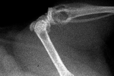
This X-ray image shows metastatic bone tumors in a mouse with kidney cancer. The metastatic tumors appear as hollow or dark areas in the bones.
Metastasis, the process by which some tumors spread from their site of origin to other body parts, accounts for more than nine out of ten cancer-related deaths. But scientists still know relatively little about the genes and biological processes that cause metastasis to occur — information that potentially could translate into new ways to stop cancers from reaching advanced or terminal stages.
Now a Memorial Sloan-Kettering research team has shed light on the mechanisms by which kidney cancer metastasizes to distant organs, including the lungs, bone, and brain. Published in the journal Nature Medicine in December, the findings point to potential therapeutic strategies to control the spread of the disease. The study also offers scientific insights that could advance metastasis research in other cancer types.

Focusing on Kidney Cancer

The research was led by cancer biologist Joan Massagué, Chair of the Sloan-Kettering Institute’s Cancer Biology and Genetics Program and Director of the Metastasis Research Center. In recent years, his laboratory has uncovered basic causes of metastasis in a number of cancer types, including breast and lung cancers.
“In this study, we focused on clear cell renal cell carcinoma, the most common subtype of kidney cancer, for which there is an urgent need for more-effective therapies,” says postdoctoral fellow Sakari Vanharanta, the first author of the Nature Medicine report. “The metastatic form of this disease is almost always incurable.”
In the vast majority of patients, clear cell renal cell carcinoma (ccRCC) tumors carry DNA changes in a gene called VHL. These mutations have been shown to cause the formation of primary kidney tumors, but they do not necessarily lead to metastasis. Until recently, researchers did not know what makes some renal cell carcinoma cells capable of forming secondary tumors in distant organs.

New Potential Drug Targets

In the recent study, the team addressed this question by performing experiments in mouse models and cell lines, and by analyzing biological and clinical data from more than 700 patients with ccRCC, whose tumors had been analyzed in large-scale cancer genomics projects.
They discovered that two genes called CYTIP and CXCR4 are activated in metastatic tumor cells but inactive in non-metastatic cells. Their experiments suggest that the activation of the two genes might be essential for the spread of kidney cancer.
CXCR4 has been linked to metastasis before in this and other tumor types, including breast cancer,” Dr. Vanharanta says. “Now, our study shows that blocking CXCR4 function with a drug called plerixafor can reduce kidney cancer metastasis in mice.” Plerixafor (MozobilTM) is currently used to stimulate blood stem cells in some cancer patients treated by bone marrow transplantation.
The researchers plan to investigate further whether CXCR4 and CYTIP, and other genes identified in the study, might offer new targets for the development of more-effective drugs for kidney cancer.

Exploring the Epigenetics of Metastasis

In addition, the investigators explored the mechanisms by which the CXCR4 and CYTIP genes are switched on in kidney cancer cells to incite metastasis. Their study revealed that the genes undergo a series of epigenetic changes — modifications in the proteins that package a cell’s DNA and regulate genes.
Unlike gene mutations, which alter a cell’s genetic code, epigenetic changes leave the DNA sequence unaffected. Nevertheless, such changes can influence a cell’s behavior by switching individual genes on or off.
Epigenetic modifications are commonly seen in many types of cancer and have recently been associated with more-advanced disease. However, little is known about the specific genes and mechanisms by which tumor cells may reconfigure their epigenetic makeup, causing a person’s disease to progress and establish itself in new organs.
“Our study has demonstrated with clear examples how epigenetic alterations can lead to the activation of metastasis-inducing genes,” Dr. Vanharanta notes. “This is a conceptual advancement that is likely to help us understand how metastasis occurs in kidney cancer as well as in other cancer types.”

Comments

Congratulations!!!! to Director Joan Massague and PostDoc Sakari Vanharanta. This text was more understandable for me. Thank You!
Super research!! Congrats much!
This is what we patients truly need: new approaches, brave research, quick human tests! Thank you for your brilliant work! We live in hope.
Congratulations to Director Joan Massague and PostDoc Sakari Vanharanta!! I am a survivor of Renal Cell Carcinoma, which had matastised in my lung, aorta and liver through a clinical trial conducted by Dr. Motzer in 2004-2005 at MSKCC. It has been since then that I have been cured and survived through the clinical trial that gave me Pegalated Interferon. I am also a donor to MSKCC and am glad to see the research projects providing great results. Keep it up, you guys out there can do it and we can soon win the battles against cancer. God Bless!
Congrats to Drs on this valuable research. I am suffering from mRcc with lungs and bones having metastasis. I hope new medicine will come before I die. Sutent and Afinator have failed on me. Any valued advice for my survival will be appreciated. I have also shown to Dr James Heish of Sloan Memorial.
You have successfully blocked the gene CXCR4 in your laboratory mice to prevent metastasis. I have removed the kidney with ccRCC, stage III pT3a four and half years ago and no clear evidence of metastasis has been found as yet in 6 monthly follow up CT scans. I wish, my RCC is the same type as that of the mice and I would be glad to become a mice for a limited period of time !

Add a Comment


Your e-mail address is kept private and will not be shown publicly.


CPRIT FAILURES

What exactly came out of the CPRIT experience so far and, we said this before, IT IS
lack of Vision, baldness and authenticity.   It was conformity to existing systems and some greed!
CPRIT was taken over by University fund raising people who spent more time imagining ways to funnel money to Universities without asking those Universities to deliver something CPRIT should have wanted.  Without definition of what was wanted by CPRIT, without a vision from CPRIT, there is no way to calibrate and measure University output.   Limiting the number of research applications by university should not have become the only control mechanism of waste at CPRIT.  How these research topics are relevant to the master plan at CPRIT and how much they will advance us toward cancer cure should have been the compass!     How they fit into the plan and vision at CPRIT.  It was shocking to me to hear the scientific leader at CPRIT saying she will not sit in meetings where funding will actually be attributed to avoid the ongoing fight among universities.  Who is watching the plan for cancer cure if any at CPRIT!  Once one has gotten the power it is time to shine by baldness and resistance to local undue pressures.  Rise above politics, do not bend to pressure because it may skew your outcome.  And clearly only the truth wins.  Carry yourself true to the master plan because it is your only true friend and Compass.  Religious people believe in the truth of the Gospel and not in the cloth and power of the pastor.  And a Pastor is good when his words stick to the bible.  Many pastors have been saved from politics within the church because the community support them, the community believe they speak the truth of the bible!

Those of us who have worked in mass implementation of public health program recognize the power of ANIMATORS in the adoption process of healthy behaviors.  Dear ABBY is an example.  Prevention programs need a GURU or two in Texas.   Allowing Survivorship programs to be disseminated in 4 or 5 locations throughout the state to acquire and develop such persona is critical.  People will not comply until they are convinced by messages from their leader/GURU.  Not everybody of course listen to a guru in a free society but those who will, represent a gain for our society as long as the message is improving health behavior.  2 months ago, CPRIT was still looking at how success will look like.

When it comes to search for a cure, it is an open race.  To send 42% of funds to one institution has no justification whatsoever.  It is a demonstration of being under the thumb and nothing else.  There is a miss opportunity here of grave consequences.  CPRIT should do like most big organization, regional offices with real power to give fund based on local needs and local constraints. This the best way to address the variety of population needs in Texas, and limit disparity variation in assistance to communities.  This concentration of power in Houston or Austin is not warranted and increase to susceptibility to political influence.  Regional Komen foundation offices have dominion over local programs.  Check with them!  Their central office still control what is going on.  But this way over 60% of tax money will not end up in one city (HOUSTON) as it is the case now at CPRIT.

let's give to CPRIT a better chance, a state dimension, people needs to know what is CPRIT.  When we speak to our colleagues about CPRIT, many oncologists look at us as if we dropped from the moon. Only a handful of doctors have heard of it!  CPRIT is a secret known only to few researchers.  And Universities want to keep it that way. Some offices are now coming to CRBCM to train on how to best access CPRIT portal for future applications,  Come on guys, 3 billions and you don't want people to know about!  Even Medscape wrote a piece about CPRIT only after the CPRIT debacle happened!

who has the vision for the cure?
who has the  baldness to create the new state wide CPRIT?
who will resist the power of the networking?
who wants all the people of Texas to get involved?
THAT IS THE LEADER NEEDED AT CPRIT!
DIFFERENTIATION AND CANCER CURE II

Since our primary objective is to find "natural ways" for the cure.  How does differentiation fit into the strategy.  We have described how Differentiation is linked to suppression or silencing of certain genes (WT1), and over-expression of other (specific transcription factors or types of RAS).  The cell under differentiation shows us how to do it to control gene expression (various pattern of Methylation).  Even the location of the differentiation Vs another location where this differentiation does not occur, gives us local environmental conditions prohibiting such differentiation (why wings on the back and not the front).  What environment makes the cell tick is important in Hematologic cancers.

The other opportunity that differentiation offers is that it bring natural cell death to even the cancer cell.  Indeed full differentiation renders the cell susceptible to turn over mechanisms which includes Apoptosis.
The notion that amount of methylation is important for cell survival bring an interesting notion that we should be quantifying this parameter and registering pattern to differentiate cancer cells from normal cell. and that selective demethylation could be curative.

When you block differentiation, specific tissue development may be compromised all together. In such formed tissue, could blocking these tissue differentiation molecules affect more proliferating cells? Certain differentiation (Mucin production) are protective for the cancer (TIM-3).  Silencing or antibody receptor to related growth factor allow for a weaker cancer.   Can we switch differentiation of cancer cells into easier cancer to treat through manipulation of growth factor or alteration of splicing sequence or quantitative or qualitative change of transcription factors.  By gene Knock out or silencing, can we either commit cells to full differentiation or remove cells ability to go back to a proliferative state....

Genes of Differentiation
WT 1
OCT4
SOX 4
KLF4
MYC
NANOG
PRC2
WINT
STAT3
NGF
Variable genes
RAS
CK5,6
YB-1
Nomenclature comment to follow

Friday, February 1, 2013

Mayo Study: Common Diabetes Drug May Treat Ovarian Cancer

Sunday, December 02, 2012
Diabetic patients with ovarian cancer who took the drug metformin for their diabetes had a better survival rate than patients who did not take it, a study headed by Mayo Clinic shows. The findings, published early online in the journal Cancer, may play an important role for researchers as they study the use of existing medications to treat different or new diseases.
Metformin is a widely prescribed drug to treat diabetes, and previous research by others has shown its promise for other cancers. The Mayo-led study adds ovarian cancer to the list.
Researchers compared the survival of 61 patients with ovarian cancer taking metformin and 178 patients who were not taking metformin. Sixty-seven percent of the patients who took metformin were surviving after five years, compared with 47 percent of those who did not take the medication. When the researchers analyzed factors such as the patients' body mass index, the severity of the cancer, type of chemotherapy and quality of surgery, they found that patients taking metformin were nearly four times likelier to survive, compared with those not taking the medication.
"Our study demonstrated improved survival in women with ovarian cancer that were taking metformin," says co-author Sanjeev Kumar, M.B.B.S., a Mayo Clinic gynecologic oncology fellow. "The results are encouraging, but as with any retrospective study, many factors cannot be controlled for us to say if there is a direct cause and effect. Rather, this is further human evidence for a potential beneficial effect of a commonly used drug which is relatively safe in humans. These findings should provide impetus for prospective clinical trials in ovarian cancer."
The results may pave the way for using metformin in large-scale randomized trials in ovarian cancer, researchers say. Given the high mortality rate of ovarian cancer, researchers say there is a great need to develop new therapies for ovarian cancer. Metformin may potentially be one of these options.
Other study authors are Alexandra Meuter, M.D., of Ludwig-Maximilians University, Munich, Germany; Shailendra Giri, Ph.D., and Ramandeep Rattan, Ph.D., of Henry Ford Health System; Jeremy Chien, Ph.D., of the University of Kansas Medical Center; Prabin Thapa, M.S.; Carrie Langstraat, M.D.; William Cliby, M.D.; and Viji Shridhar, Ph.D. of Mayo Clinic.
This work was supported, in whole or in part, by Fred C. and Katherine B. Andersen Foundation grant, and CA123249 and P50 CA136393 National Institutes of Health grants.
###

About Mayo Clinic

Mayo Clinic is a nonprofit worldwide leader in medical care, research and education for people from all walks of life. For more information, visit MayoClinic.com or MayoClinic.org/news.
Journalists can become a member of the Mayo Clinic News Network for the latest health, science and research news and access to video, audio, text and graphic elements that can be downloaded or embedded.

Media Contact

Kelley Luckstein
507-284-5005 (days)
507-284-2511 (evenings)
newsbureau@mayo.edu

REVIEW ARTICLE 

Can Metastatic Colorectal Cancer Be Cured?

By David L. Bartlett, MD1,3, Edward Chu, MD2,3 | March 13, 2012
1 Division of Surgical Oncology, Department of Surgery 2 Division of Hematology-Oncology, Department of Medicine and Pharmacology & Chemical Biology 3 Molecular Therapeutics Drug Discovery Research Program, University of Pittsburgh Cancer Institute, University of Pittsburgh School of Medicine, Pittsburgh, Pennsylvania


ABSTRACT: Significant advances have been made in the treatment of metastatic colorectal cancer (mCRC). Development of the targeted biologic agents and their integration with cytotoxic chemotherapy regimens has led to improvements in clinical efficacy. Despite these gains, the overall impact of these combination regimens in mCRC therapy has been relatively modest. While 2-year survival has improved, substantive gains have yet to be made in 5-year survival. However, a small subset of patients can be cured of their metastatic disease, with prolonged 5- and 10-year overall survival. This select group of patients includes those with metastatic disease limited to the liver or other organ-specific sites, as these patients are able to undergo surgical resection at the time of diagnosis or following conversion therapy with the appropriate integration of chemotherapy. A multimodality team-based approach involving medical oncologists, surgical oncologists, radiologists, and other healthcare providers is absolutely critical for the success of this therapeutic approach. This article reviews the main issues that must be considered from the surgical oncology and medical oncology perspectives, respectively.

Introduction

In 2012, colorectal cancer (CRC) continues to be a major public health problem. In the United States this year, there will be an estimated 147,000 new cases diagnosed and nearly 50,000 deaths resulting from this disease.[1] Worldwide, approximately 1 million new cases of CRC are diagnosed each year, with nearly 500,000 deaths attributed to this disease annually. About 25% of patients present with metastatic disease, and of this group, 50% to 75% will have disease confined to the liver.[2-4] In patients who present initially with early-stage disease, up to 50% will eventually develop metastatic disease, with the liver being the most common site. Another 10% to 20% of patients will present with disease involving the lung and other less common sites of metastatic involvement, including the peritoneum, ovaries, adrenal glands, bone, and brain.[5,6]
(MORE: Metastatic Colorectal Cancer: Potential for Cure?)

When metastatic disease is limited to an organ-specific site, an important consideration is whether the disease is resectable at the time of initial diagnosis or whether it is initially deemed to be unresectable but may become resectable with the up-front use of chemotherapy. With the integration of chemotherapy and surgical resection, overall 5-year survival rates on the order of 30% to 40% can now be achieved. A multidisciplinary, team-based approach involving surgeons, medical oncologists, radiologists, and other healthcare professionals is required to determine the optimal timing and sequence of surgery and chemotherapy.
This article reviews the multidisciplinary approach to patients who have organ-limited metastatic CRC (mCRC), with the main focus being on liver- limited disease. In particular, the surgical and chemotherapy aspects of disease management will be discussed.

Surgical Considerations for Patients With Metastatic Disease

Historically, the setting of liver-limited metastases from CRC has been one of the few examples of curative metastasectomy in oncology. Even before the development of effective chemotherapy agents, surgical resection of limited hepatic metastases was associated with prolonged survival and cures.[7] Several important prognostic factors, such as disease-free interval, number and size of metastases, presence of extrahepatic disease, and stage of the primary cancer, have all helped to define the expected cure rate for hepatic metastasectomy. For patients with metastases defined by the most favorable prognostic categories, cure rates of 24% have been achieved with surgery alone.[8] The indications for surgical metastasectomy were for patients with disease limited to the liver, a total of four or fewer metastases, unilobar involvement, tumors of less than 5 cm in their greatest diameter, and a disease-free interval of at least 6 months.[9-12] It is, therefore, not surprising that the development of more effective chemotherapy has led to a significant improvement in overall survival and cure rates, as well as an expansion of the indication for metastasectomy. This indication has evolved into resection of any disease that allows for adequate hepatic residual volume for liver regeneration and survival, assuming there has been a response to neoadjuvant chemotherapy.[13] In the past, surgeons were appropriately concerned that resection of visible disease would be followed by rapid recurrence from microscopic metastases in the residual liver. However, incorporation of effective neoadjuvant and/or conversion chemotherapy, as will be discussed in this article, provides greater confidence that micrometastatic disease can be eliminated and that removal of gross disease can lead to long-term cure. In addition, as hepatic surgery has become safer and easier for the patient, there is now wider acceptance of incorporating hepatic resection into a multimodality strategy to prolong survival.
The options for local and regional treatment of hepatic metastases have become broad, and include surgical resection, local ablation therapy, hepatic arterial infusion therapy, transarterial chemoembolization, radiomicrosphere therapy, and isolated hepatic perfusion.[14,15] Each of these approaches has been associated with long-term cures, although surgical resection and local ablation strategies have been the most effective. The goal for surgical resection is to achieve a negative microscopic margin. Given the concern about microscopic extension beyond the visible tumor, a 1-cm margin around the tumor is ideal. Numerous coagulation devices exist to enhance the safety of parenchymal transection by limiting blood loss. Minimally invasive approaches, such as laparoscopic and robotic assistance, have become commonplace, and they are associated with reduced blood loss, shortened hospital stay, and decreased narcotic usage postoperatively.[16,17] For patients undergoing multimodality therapies, minimally invasive surgery may also improve quality of life during treatment and decrease the recovery time necessary before adjuvant chemotherapy is administered. The options for resection include extended lobectomy, lobectomy, segmentectomies, and nonanatomic wedge resections. Many surgeons remove the least amount of liver tissue feasible to preserve the anatomy for future resections, if necessary, while others prefer formal anatomic resections in order to provide the best chance of a negative margin. These two approaches have not been directly compared in a randomized trial; however, retrospective data suggest that the ability to achieve a negative margin, as opposed to the specific type of resection, determines long-term prognosis.[18]
Local ablative approaches have provided an alternative to surgical resection for patients with mCRC. These approaches include radiofrequency ablation (RFA), microwave ablation, cryotherapy, and focused radiotherapy (eg, using the CyberKnife). RFA is a reliable technique to ablate metastases up to 5 cm in size. However, it has limited efficacy in centrally located tumors in which proximity to the main portal triads or hepatic veins may cause bile duct injury, extensive hepatic necrosis, or inadequate tumor cell death adjacent to the vessels. The potential advantages of these local strategies over surgical resection include enhanced safety, outpatient percutaneous treatment options, and the ability to preserve hepatic parenchyma. The local recurrence rate after local ablative procedures is clearly higher than with surgical resection, with rates as high as 34% having been reported.[19] The local recurrence rate at the site of ablation is influenced by the size and location of the metastatic lesions, as well as the use of percutaneous vs laparoscopic approaches. Although local recurrence can often be salvaged with repeat ablation or resection, for patients with limited comorbidities in whom the goal is curative intent, surgical resection is the preferred and most reliable method for actual cure. A meta-analysis of nonrandomized studies comparing RFA with surgical resection demonstrated an improvement in 5-year survival for patients treated with hepatic resection.[20]
TABLE 1

Prognostic Factors for Cure After Surgical Resection
The curative potential of surgical resection for hepatic metastases from CRC varies depending on a number of important prognostic factors (Table 1). Nomograms for predicting cancer-related survival have been developed, and may be helpful when considering the utility of resection.[21] A patient’s risk for morbidity and mortality also plays a significant role in defining the eventual treatment strategy. Surgical resection is still associated with a defined mortality rate of 2.8% (0 to 6.6%), which is influenced, in large part, by the health of the background liver.[22,23] Liver failure is the most common cause of death after hepatectomy, and as discussed below, this complication is influenced by the specific type and cumulative dose of chemotherapy received. The indications for surgical resection are currently based on feasibility and safety in patients who have responded to chemotherapy. It is critically important for the surgical resection to leave 20% to 25% of functioning liver volume (future liver remnant [FLR]) in patients with a normal background liver, and 40% of liver volume in patients whose background liver is diseased from previous chemotherapy.[24] Preoperative planning CT scans, including residual volume calculations, are essential when planning an extended or bilobar resection.[25]
To date, more than 750 series of hepatic metastasectomy for metastatic CRC have been reported in the literature. The actuarial 5-year survival rate for patients who underwent R0 resections (negative margins) was 30% when combining 16 well-reported series of more than 100 patients with follow-up greater than 2 years (15% to 67%).[22] While 5-year survival was historically considered a cure for this disease, because of advances in systemic chemotherapy an increasing number of patients are now living with their disease beyond 5 years. A single-institution study of 455 patients revealed a median overall survival of 33 months, with 5- and 10-year actuarial survival rates of 34% and 25%, respectively.[26] In that study, 124 patients were identified as actual 5-year survivors (27%), and of this group 59 were found to be 10-year survivors. This finding suggests ongoing disease-related mortality beyond the 5-year time-frame, with actual cure rates of 10% to 15%. Randomized clinical data suggest an improvement in disease-free survival when systemic chemotherapy is incorporated as part of a combined neoadjuvant and postoperative adjuvant approach, as will be discussed in detail in this article.
With the extended indications for hepatic metastasectomy in the presence of active systemic chemotherapy, larger resections can now be safely and effectively performed. Commonly used techniques include staged resections for bilobar disease and preoperative portal vein occlusion to achieve compensatory hypertrophy and safer extended resections.[27,28] While there appear to be impressive actuarial 5-year survival rates in these series of extensive surgical resections, it is expected that the true cure rate will be much lower. When looking at patients with initially unresectable colorectal liver metastases who were treated with chemotherapy and then resected, 16% of this group were considered cured, with a disease-free interval of more than 5 years after metastasectomy.[29] On multivariate analysis, the main predictors of cure included maximum size less than 3 cm, no more than three metastatic lesions, and complete pathologic response.
Pages: 1  2  3  


Systemic Chemotherapy

Long-term cures are exceedingly rare when patients with organ-limited mCRC are treated with chemotherapy alone. In a retrospective review of 2751 patients with metastatic CRC, during a median follow-up of 10.3 years, only 6 (0.24%) were found to be free of disease after having received chemotherapy alone.[30] It is now well established that a multimodality strategy results in a much higher chance of long-term cure. In patients with organ-limited disease, chemotherapy is administered in three main settings, which include neoadjuvant therapy, conversion therapy, and adjuvant therapy. Neoadjuvant therapy refers to chemotherapy given to patients with potentially resectable disease, while conversion therapy refers to chemotherapy given to patients deemed to have initially unresectable disease. Adjuvant chemotherapy is use of chemotherapy following an R0 surgical resection, with the intent of preventing disease recurrence.

Neoadjuvant Chemotherapy

Up to 20% to 30% of patients with liver-limited mCRC may have potentially resectable disease at the time of initial presentation. However, because a large proportion of patients experience recurrence of their disease either in the liver or systemically, chemotherapy has been integrated in their up-front care to improve upon the potential benefit of surgery.
(MORE: Metastatic Colorectal Cancer: Potential for Cure?)

Several clinical trials have specifically evaluated the role of neoadjuvant therapy for patients with potentially resectable liver metastases. In a single-arm trial involving 20 patients, neoadjuvant therapy with a weekly administration of FOLFOX (fluorouracil [5-FU], leucovorin/folinic acid [LV], and oxaliplatin(Drug information on oxaliplatin) [Eloxatin]) resulted in a partial or complete response in all patients enrolled.[31] A total of 16 patients underwent a potentially curative resection, with 7 developing recurrence during the median follow-up period of 23 months. A phase II trial of neoadjuvant therapy investigated bevacizumab(Drug information on bevacizumab) (Avastin) plus CapOx, the combination of capecitabine(Drug information on capecitabine) (Xeloda) and oxaliplatin.[32] In this study, 56 patients received 6 cycles of therapy prior to surgical resection, and a remarkably high objective response rate of 73% was observed. A total of 52 of the 56 patients were able to undergo an R0 resection, with complete pathologic response occurring in nearly 10% of patients. Given concerns over the potential risks of bleeding or wound-healing complications, bevacizumab was not given with the last cycle of chemotherapy prior to surgery. This study is important as it showed that bevacizumab could be safely administered to patients with no increased risk of intraoperative bleeding or wound-healing complications. Moreover, it was estimated that normal liver regeneration occurred in all but one patient.
The European Organisation for Research and Treatment of Cancer (EORTC) randomized phase III trial 40983 investigated use of perioperative FOLFOX4 chemotherapy in patients with up to four resectable liver metastases. In this study, patients were randomized to surgery alone or to receive 6 cycles of FOLFOX4 before surgery and 6 cycles of FOLFOX4 after surgery.[33] The overall response rate was 43% in patients receiving chemotherapy. Of note, surgery was performed in 83% of patients randomized to chemotherapy and in 84% of patients randomized to surgery alone, providing evidence that use of initial chemotherapy did not compromise the ability of patients to undergo surgical resection. While there was an increased risk of postoperative complications in patients receiving neoadjuvant chemotherapy, these events were reversible and not associated with an increased risk of mortality. When the entire group of randomized patients was considered, a 7.3% increase in progression-free survival (PFS) at 3 years was observed in patients receiving chemotherapy, although this difference did not reach statistical significance. However, in the group of patients who underwent surgical resection, a significant 9.2% improvement in 3-year PFS was, in fact, observed.
Adam et al examined the influence of the response to neoadjuvant chemotherapy on the eventual outcome in patients following surgical resection of multiple liver metastases.[34] In this retrospective analysis of 131 patients, 44% underwent hepatectomy after achieving an objective tumor response, 30% went to surgical resection after tumor stabilization, and 26% were surgically resected after tumor progression. Five-year survival was significantly lower in the group of patients who had evidence of tumor progression, compared with patients who had evidence of tumor response (8% vs 37%). Of note, patients with stable disease on neoadjuvant chemotherapy had only a slightly worse prognosis with respect to 5-year survival, compared with responders (30% vs 37%). Disease-free survival in patients who progressed on neoadjuvant chemotherapy was only 3%, compared with rates of 21% and 20% for patients with tumor response or stable disease, respectively. Based on this study, it is clear that tumor progression before surgery is associated with extremely poor clinical outcome, and in this setting, hepatic resection should be avoided in patients who are deemed to be nonresponders to preoperative chemotherapy.
Neoadjuvant chemotherapy may be associated with complete disappearance of some or all of the hepatic metastases on imaging studies (approximately 18% of tumors will disappear completely).[35] Pathological complete response is associated with a high rate of long-term cure after surgical resection (5-year survival of 79%).[36] Controversy exists regarding the need to resect patients with complete radiographic responses, to achieve long-term cure. Up to 70% of these sites of complete radiographic response are associated with pathologic complete response or failure to recur at these sites.[36,37] The remaining 30% of patients are at risk of disease recurrence if resection is not performed. Thus, curative therapy should include resection of these regions, although the potential risk of disease recurrence at other sites must also be taken into consideration.

Conversion Therapy

The majority of patients will present with liver metastases from CRC that are unresectable or not optimally resectable based on their size, number, or location at the time of initial assessment. In this setting, conversion therapy is used in appropriately selected patients. The primary focus, therefore, is on achieving downsizing of the metastatic disease that is sufficient to allow surgical resection to be performed, but not with the goal of achieving a complete or even maximal response.
Adam and colleagues in France have had the largest experience in this area to date, and their work has provided important insights into the potential role of conversion therapy.[38-40] In their original series of 701 patients with initially unresectable liver metastases, treatment with oxaliplatin-based chemotherapy resulted in downsizing in nearly 15% of patients, and subsequent surgery. Based on 5-year follow-up after surgery, 22% of patients had no evidence of residual or recurrent disease. When stratified according to the underlying reasons for initial unresectability, the 5-year overall survival (OS) rates were 60% for patients with large tumors, 49% for those with poorly located tumors, and 34% for patients with multinodular tumors. In an expanded series of 1439 patients treated with a broader range of cytotoxic chemotherapy, the conversion rate was 12.5%, with a 5-year survival rate of 33%.
Folprecht and colleagues[41] conducted an interesting analysis of all published/presented clinical trials and retrospective studies of the rate of objective response and the subsequent rate of resection of initially unresectable metastases. They observed a strong correlation (r = 0.96) between response rates and the subsequent resection rate in patients with isolated liver disease. Moreover, their analysis confirmed that patient selection and efficacy of preoperative chemotherapy were strong predictors of potential resectability of liver metastases. Since this analysis, several prospective clinical trials incorporating systemic chemotherapy plus surgery have been performed. In these studies, use of oxaliplatin- vs irinotecan(Drug information on irinotecan)-based chemotherapy has shown similar clinical outcomes.[42,43] Of note, approximately 20% to 30% of patients were able to undergo R0 surgical resection. Two trials have directly compared the clinical efficacy of FOLFOX plus irinotecan (FOLFOXIRI), an aggressive regimen that incorporates the three active cytotoxic agents, against that of FOLFIRI (5-FU, LV, irinotecan). Falcone et al randomized patients with mCRC to receive either FOLFOXIRI or FOLFIRI, and they reported a significant increase in R0 resection for the subgroup of patients with liver-only metastases who were randomized to the FOLFOXIRI arm.[44] The R0 resection rate was 36% in the FOLFOXIRI arm vs 12% in the FOLFIRI arm (P = .017). Despite the increased clinical activity of FOLFOXIRI, patients receiving this regimen experienced a significantly higher incidence of grade 3/4 toxicity in the form of myelosuppression and neurotoxicity. In contrast to the positive findings of the Falcone study, Souglakos et al observed a nonsignificant increase in overall response rate (43% vs 33.6%), conversion rate (10% vs 3.4%), and R0 resection rate (8.8% vs 3.4%).[45] A pooled analysis of the Falcone phase III study and two phase II studies reported an overall response rate of 70% with the FOLFOXIRI regimen and a 19% R0 resection rate. The 5-year disease-free survival (DFS) and OS were 29% and 42%, respectively.[46]
TABLE 2

Select Trials Reporting Conversion of Unresectable Metastatic CRC to Resectable Metastatic Disease
Is there an optimal cytotoxic chemotherapy regimen for conversion therapy? To date, there has been a significant absence of randomized trials directly comparing the various chemotherapy regimens in patients with liver-limited disease. In reviewing the literature, it appears that irinotecan- and oxaliplatin-based regimens yield approximately the same rate of conversion, on the order of 20% to 30%. While FOLFOXIRI appears to result in higher conversion rates, in the 40% to 60% range, and higher R0 surgical resections, this treatment regimen is clearly associated with increased toxicity and should be used only in certain select patient populations. Upon review of the recent National Comprehensive Cancer Institute (NCCN) guidelines, several regimens are currently recommended, and they include FOLFIRI, FOLFOX, CapOx, and FOLFOXIRI.[47]
The introduction of targeted therapies with either the antiangiogenic agent bevacizumab or the epidermal growth factor receptor (EGFR) inhibitors cetuximab(Drug information on cetuximab) (Erbitux) and panitumumab (Vectibix) has improved the clinical efficacy of chemotherapy in patients with mCRC. As a result, combination regimens incorporating these agents have now been evaluated in clinical trials for patients with liver-limited metastases.
The addition of the anti–vascular endothelial growth factor (VEGF) antibody bevacizumab to either FOLFOX or to capecitabine and oxaliplatin (XELOX/CapOx) vs the cytotoxic chemotherapy regimens alone was investigated in a randomized phase III trial in advanced mCRC.[48] Unfortunately, there was only a slightly higher incidence of R0 surgical resection with bevacizumab (8.4%) vs chemotherapy alone (6.1%).
The anti-EGFR antibodies cetuximab and panitumumab have been approved for use in patients with mCRC.[49] Subsequent studies have shown that these agents are active only in patients with wild-type KRAS tumors. KRAS mutations occur in up to 30% to 40% of patients with CRC, and they typically involve codon 12 or 13. In general, KRAS mutations lead to resistance to antibody therapy. However, recent studies have suggested that the G13D mutation in codon 13 may still allow for sensitivity to anti-EGFR antibody therapy, in sharp contrast to mutations in codon 12.
Retrospective analyses of clinical trials in mCRC have provided insights into the potential role of cetuximab in the treatment of liver-limited disease. In a phase II trial of FOLFOX plus cetuximab, 37 of the 43 patients enrolled had liver involvement, and in 17 of these patients, the liver was the only site of metastatic disease.[50] An objective response was seen in 34 of the 37 patients; 10 of these patients underwent surgical resection of their metastases, including 8 patients with liver metastases. In a series of 151 patients with unresectable mCRC liver metastases refractory to systemic chemotherapy, the addition of cetuximab to combination chemotherapy allowed 27 patients to undergo surgical resection, and of this group, 25 underwent potentially curative hepatectomy.[51] Of note, this group included a majority of patients who were deemed to have either technically unresectable or marginally resectable disease. Moreover, the incorporation of cetuximab with chemotherapy conferred significant clinical benefit, with median progression-free survival (PFS) and OS of 13 and 20 months, respectively.
Several single-arm phase II trials have investigated the combination of cetuximab with either irinotecan- or oxaliplatin-based regimens. Min et al reported a radiologic response rate of 39%, with 30% of patients treated with FOLFIRI plus cetuximab able to undergo resection of their liver metastases.[52] Nearly identical results were observed with the combination of FOLFOX and cetuximab, which yielded an R0 resection rate of 29%.[53]
Two recent randomized studies have investigated the safety and efficacy of cetuximab in combination with either FOLFIRI[54] or FOLFOX.[55] The addition of cetuximab to FOLFIRI significantly increased the overall response rate (59% vs 43%; P = .004) in patients with wild-type KRAS when compared with FOLFIRI alone, and this resulted in a higher number of patients able to undergo R0 surgical resection (4.3% vs 1.5%). An exploratory analysis revealed a two-fold higher rate of R0 surgical resection in patients with liver-limited disease (9.8% vs 4.5%).[54] Similar findings were reported by Bokemeyer et al[55] with the combination of cetuximab plus FOLFOX4. The overall response rate increased from 37% to 61% in patients with wild-type KRAS and in those treated with the combination vs FOLFOX4 alone. This improvement in response rate in patients treated with the combination was associated with an increase in the R0 resection rate from 2.4% to 4.7%.
A trial of 114 patients with initially nonresectable liver-limited metastases randomized patients to receive cetuximab in combination with either FOLFOX6 or FOLFIRI. R0 resection rates of 38% and 30% were observed, respectively, with an overall R0 resection rate of 34%.[56] In a retrospective analysis of response according to KRAS status with the two arms of the trial combined, the clinical response rate in patients with wild-type KRAS was 70% compared with 41% for those with mutant KRAS. This study provides further evidence of the strong association between high tumor response rate and increased rate of liver metastasectomy.
PRIME (the Panitumumab Ran-domized Trial in Combination With Chemotherapy for Metastatic Colorectal Cancer to Determine Efficacy) was designed to evaluate the efficacy and safety of panitumumab plus FOLFOX4 vs FOLFOX4 alone as initial treatment for mCRC. The addition of panitumumab to FOLFOX4 chemotherapy significantly improved the overall response rate (57% vs 48%; P = .02) and median PFS in patients with wild-type KRAS tumors (9.6 vs 8.0 months; P = 0.01), which translated into a nonsignificant increase in median OS from 19.7 to 23.9 months.
In terms of surgical resection, metastasectomy of any site was attempted in 10.5% of patients treated with the combination regimen as opposed to 9.4% of patients treated with chemotherapy alone. However, the R0 resection rate was higher in patients with wild-type KRAS tumors and liver-limited disease (28% vs 18%) who were treated with panitumumab plus FOLFOX4. At the time of the most recent analysis, median OS had not been reached in patients who underwent R0 liver resection, in contrast to a median OS of 23.6 months in those who were unable to undergo complete surgical resection.[57]
Pages: 1  2  3


Pages: 1  2  3  
REVIEW ARTICLE 

Can Metastatic Colorectal Cancer Be Cured?

By David L. Bartlett, MD1,3, Edward Chu, MD2,3 | March 13, 2012
1 Division of Surgical Oncology, Department of Surgery 2 Division of Hematology-Oncology, Department of Medicine and Pharmacology & Chemical Biology 3 Molecular Therapeutics Drug Discovery Research Program, University of Pittsburgh Cancer Institute, University of Pittsburgh School of Medicine, Pittsburgh, Pennsylvania


Adjuvant Chemotherapy

To date, only a limited number of clinical trials have investigated the role of adjuvant chemotherapy following surgical resection of organ-limited metastases. Two randomized phase III trials were conducted to determine the potential role of adjuvant chemotherapy with 5-FU/ LV vs surgery alone.[58,59] Both trials showed a nonsignificant trend for improvement in DFS. Unfortunately, both studies closed prematurely due to slow patient enrollment. As a result, neither study had sufficient statistical power to demonstrate the predefined difference in OS. A pooled analysis of the individual data from these two trials was subsequently conducted by Mitry et al to improve the statistical power of the survival analysis. This analysis showed a marginally significant trend toward improved progression-free survival for patients receiving chemotherapy (27.9 vs 18.8 months).[60] This study is important as it provides proof of concept for the potential role of adjuvant chemotherapy in patients who have undergone curative resection of liver or lung metastatic disease. Unfortunately, a randomized phase III trial was unable to document the benefit of the FOLFIRI regimen as adjuvant therapy following surgical resection of liver metastases when compared with infusional 5-FU/LV.[61]
What should the recommendations be for adjuvant chemotherapy following surgical resection? Although definitive clinical data are lacking, the current approach would be to offer adjuvant therapy with an oxaliplatin(Drug information on oxaliplatin)-based regimen, whether it be FOLFOX or XELOX, for a defined 3- to 4-month period. As is the case for the adjuvant treatment of early-stage colon cancer, there is presently no role for a biologic agent, such as bevacizumab(Drug information on bevacizumab) or the anti-EGFR antibodies cetuximab(Drug information on cetuximab) and panitumumab, in oxaliplatin-based chemotherapy. Further support for this approach comes from the recently published NCCN clinical practice guidelines for adjuvant therapy of resected metastatic disease, which recommend a shortened course of cytotoxic chemotherapy, as would be offered for patients with resected stage III colon cancer. [47]

Limitations of Chemotherapy

FIGURE 1

Comparison of Survival of a Group of Patients With Colorectal Metastases to the Liver and a Second Group With Carcinomatosis
(MORE: Metastatic Colorectal Cancer: Potential for Cure?)

The role of chemotherapy is to enhance the outcomes of surgery and/or permit potentially curative resection to be performed. Unfortunately, chemotherapy has potential disadvantages, which relate to direct toxic effects on the liver, leading to an increased risk of potential postoperative complications. There is now a large body of evidence showing that systemic chemotherapy can result in nonalcoholic fatty liver disease and sinusoidal injury. The chemotherapy-associated liver disease ranges from steatosis to steatohepatitis (CASH).[62] Steatosis resulting from chemotherapy and/or any other etiology has been shown to lead to a higher rate of complications following hepatic resection. However, the development of CASH appears to hold greater significance.[63] Of note, CASH appears to be more closely associated with the use of irinotecan(Drug information on irinotecan)-based chemotherapy and to occur more commonly in patients with higher body mass index.[64] The development of CASH has been associated with a higher postoperative mortality rate related primarily to postoperative liver failure. In one series, the 90-day mortality rate in patients with steatohepatitis was 14.7% vs 1.6% for those who did not have steatohepatitis.[65] In contrast to treatment with irinotecan, oxaliplatin-based chemotherapy has been typically associated with liver sinusoidal injury.[62,65,66] In more severe cases, perisinusoidal fibrosis, sinusoidal obstruction, and portal hypertension have been observed. In contrast to CASH, the development of sinusoidal dilation has not been associated with an increased risk of perioperative morbidity and mortality.[67,68]

Peritoneal Carcinomatosis

While this review has focused on liver-limited metastatic disease, cures have also been reported after pulmonary metastasectomy, isolated nodal recurrences, and ovarian metastases.[69-71] While these are highly selected cases, they are worthy of consideration for patients with favorable tumor biology and/or for those who are responsive to chemotherapy. A growing field of interest has been the surgical management of peritoneal metastases from CRC, using cytoreductive surgery and intraoperative chemoperfusion with mitomycin(Drug information on mitomycin) C or oxaliplatin, combined with hyperthermia (HIPEC).[72,73] This interest stems from early randomized trials with this treatment strategy in gastric cancer and a randomized trial in mCRC from the Netherlands.[74,75] This mCRC carcinomatosis trial demonstrated an improvement in median survival in patients receiving intraoperative HIPEC, compared with systemic 5-FU/LV (22.3 months vs 12.6 months). Patients whose tumors could be completely resected from the peritoneum followed by HIPEC had an actuarial 3-year survival of 95%. A follow-up report on this trial demonstrated an overall actual 5-year survival of 45% in the HIPEC arm for patients with all disease resected.[76] A recent report from France noted a 5-year survival of 26% in patients receiving HIPEC with oxaliplatin for colorectal peritoneal carcinomatosis.[77] A number of series have compared surgical cytoreduction and HIPEC for peritoneal carcinomatosis vs surgical resection of hepatic metastases from mCRC, demonstrating similar survival curves (Figure 1).[78-80] This finding suggests that an aggressive combined-modality approach for peritoneal carcinomatosis may have a defined cure rate. Presently, most centers combine surgical cytoreduction and HIPEC with neoadjuvant and postoperative adjuvant systemic chemotherapy, such as has been described for liver-limited metastatic disease.
REFERENCE GUIDE

Therapeutic Agents
Mentioned in This Article


Bevacizumab (Avastin)
Capecitabine(Drug information on capecitabine) (Xeloda)
Cetuximab (Erbitux)
5-Fluorouracil (5-FU)
Irinotecan
Leucovorin (folinic acid)
Mitomycin C
Oxaliplatin (Eloxatin)
Panitumumab (Vectibix)


Brand names are listed in parentheses only if a drug is not available generically and is marketed as no more than two trademarked or registered products. More familiar alternative generic designations may also be included parenthetically.

Conclusions

When limited to a specific organ site, mCRC is potentially curable. To date, nearly all of the clinical studies have focused on liver-limited disease, but similar results are now being reported for patients with disease limited to the lungs, ovaries, and peritoneum. It is clear that a multidisciplinary team-based approach is required for the optimal care of this particular subset of patients. The development of an individual treatment plan comes from a careful discussion and ongoing communication among a multidisciplinary team of specialists, including surgeons, medical oncologists, and radiologists. With the appropriate integration of chemotherapy plus biological agents and surgery, up to 30% to 40% of patients with organ-limited metastatic disease can be cured. While the costs of the three biological agents—cetuximab, panitumumab, and bevacizumab—are not insignificant, the clinical evidence is now well-established that their incorporation with cytotoxic chemotherapy regimens in the neoadjuvant and conversion settings has greatly facilitated curative resection of liver-limited metastatic disease. However, further improvements are needed to enhance the clinical outcome of the remaining 60% to 70% of patients. Further refinements in whole-body and hepatic imaging should provide for a more accurate selection of the subset of patients who would benefit most from resection and would identify the presence of minimal residual disease following surgery. Finally, clinical trials are needed to develop novel cytotoxic agents and biologic/targeted agents that can be used in both the preoperative and postoperative settings to reduce the risk of local and systemic recurrence.
Financial Disclosure: The authors have no significant financial interest or other relationship with the manufacturers of any products or providers of any service mentioned in this article.
Pages: 1  2  3  


DIFFERENTIATION AND THE CURE.
5TH LAW OF NATURE

Although it would have been nice if there were clear cut cross-road between DIFFERENTIATION and PROLIFERATION.  Nature has understood that most tissues will need to be repaired particularly when it comes to the skin.  Therefore some differentiated cells could trigger de-defferentiation when repair is needed.  The question is then how could the shift occur.  What makes a hyperdifferentaiated cell shift gears and enter proliferation.
Stimuli from outside the cells including local specific growth factors (TGF Beta, nerve growth factor,Epidermal growth factor, Epiregulin, transforming Growth factor, Platelet derived GF,  and so on) attach to its cellular receptor, and trigger N,H,C, K -RAS.  and depending on the type of RAS, differentiation will be directed.  There are other point of tissue specificity but the family of RAS is one area of folk differentiation.

One other location of folk differentiation is at the level of transcription genes and at genetic  plicing  (PRPF8,
U2AF2 (keep an eye on this one),  WT-1 (looks like EGFR), PUF60, ASF/SF2,WDR,EFTUD2, PPFIA-down regulates androgen for differentiation, SF3B1 etc...

Researchers looking at NRAS and KRAS function were manipulating a Colon cancer cell when the find themself with a cell with putative stem cell features indicating that whatever stimuli being applied reversed the cell to open up totipotential characteristic.  We know that cell from skin would repair wound through proliferative process.  Increase in TNF and Interferons would yield fibrotic tissue.  After a trauma,These growth factors will direct efforts to another type of RAS.  The RAS stimulation follow the MEK transduction signal to lead to appropriate transcription and splicing factors for relevant nuclear events globally!

The thing is the more  complete the differentiation, the less the proliferative potential.

This beg a question that investigators have been struggling, forcing full differentiation by maximizing differentiation a viable strategy for cure by silencing proliferation.   One thing is for sure, high dose interferon disrupts growth factor effects and had been for a while the only treatment for Melanoma until BRAF was interfered with and now regulation of the MEK/MAP kinase has also been inhibited.  Disruption of growth factors at the membrane leads to expression of molecules and disruption of Glycocalyx susceptible to recruit
immune cells. 

THIS SAME MECHANISM would more likely lead to cirrhosis in the liver where injury lead to  change of growth factors leading to preponderance of fibrous tissue and new nodules.  This is differentiation challenged by "trauma" to the liver tissue!

GENE PROFILING, P53 STORY

P53
---------------
At least half of the Sarcomas will show activity at P53, the cellular Molecule of the year in 1993,
in up to 10% of cases, the P53 will actually be mutated. It is either overexpressed or suppressed in the rest of the cases of Sarcoma.  The P53 is suppressed if repression occurs at P14 or at CDKN2A or if MDM2 is overexpressed.  P14 shield P53 from the effect of MDM2.  P53 is also overexpressed if its degradation is stopped at the proteasome.

At the nuclear level, Acetylated P53 combines to P300 and promote P21 and Puma leading to Apoptosis.  This effect can be blocked by Fusion protein  EWS-Fli1 in Ewing Sarcoma, and by upregulation of Histone DE-Acelase 1 which effectively blocks down transcription effect of P53.

If you count right, there are 6 potential targets of interaction with P53, if you include upstream toward the membranes...calling for Multitarget therapy in those conditions where wild type P53 is overexpressed!

GENE PROFILING IS A MUST STEP IN CANCER TREATMENT!

LOOPHOLE EXISTS WHEN p53 IS BLOCKED DOWNSTREAM THOUGH,
BLOCKAGE OF p53 IN THE NUCLEAR, TRIGGERS EXPRESSION OF JAG1, HEY1 WHICH INTENSIFY NOTCH3 AND STILL LEAD TO CELL CYCLE ARREST.

MARTA Q LACY.
Professor of Medicine
Division of Hematology
Mayo Clinic College of Medicine
Rochester, Minnesota


Relapse remains a significant clinical problem for multiple myeloma. Nearly all patients eventually relapse, and, although survival rates are improving and treatment options continue to grow, the move to upfront combination therapy does limit options in relapse. FDA approved options for relapsed multiple myeloma include the immunomodulatory agents thalidomide and lenalidomide and the proteasome inhibitors, bortezomib and carfilzomib (for patients who have received at least 2 prior therapies including treatment with bortezomib and an immunomodulatory agent).
For patients not resistant or refractory to immunomodulatory agents and proteasome inhibitors, there are effective choices. However, for patients who have failed thalidomide, lenalidomide, and bortezomib survival is low.1 Disease-, regimen-, and patient-related factors contribute to therapy selection in relapse. Disease-related factors include risk as assessed by FISH and cytogenetics and duration of response to initial therapy. Patient-related factors include comorbidities, age, and performance status. Regimen-related factors include prior drug exposure, toxicity of the regimen, the mode of administration, and whether the patient has had a previous stem cell transplant. These factors should be carefully weighed when making treatment decisions for relapsed myeloma patients.
At diagnosis, Mr Johnson had no high-risk molecular markers and excellent renal function. In this setting, at Mayo, we prefer to start with lenalidomide and dexamethasone because it has high remission rates, is oral, well tolerated, and unlikely to induce peripheral neuropathy. When using lenalidomide in patients with no personal history of VTE, we favor prophylaxis with aspirin at 325 mg daily. We would use IV bisphosphonates monthly for 12 months and quarterly for 1 additional year. In patients with low-risk disease, we consider risks and benefits of maintenance therapy. If maintenance therapy is chosen, consider limiting the duration to 12-24 months. 
At relapse, if our patient had a good response and a long duration of remission (> 12 months), we favor re-introduction of the initial regimen. If the patient has suboptimal response or a short remission duration, we would change the class of drug used (eg, if initially treated with an immunomodulatory agent [thalidomide, lenalidomide], we would switch to proteasome inhibitor [bortezomib, carfilzomib]. If initially treated with a proteasome inhibitor, we would switch to an immunomodulatory agent). In this particular case, we would need to factor in that the patient now has renal failure and a new bone lesion. We generally re-introduce bisphosphonates quarterly at relapse in patients with new bone lesions. However, since this patient has renal failure, we would wait for renal improvement and favor pamidronate over zoledronic acid with a reduced dose of 30 mg (from 90 mg).1 Also due to the renal status of this patient, full-dose lenalidomide should not be used. I would favor switching to bortezomib because it can be used at full dose. However, dose-adjusted lenalidomide is also an option.2 A second ASCT may also be considered, especially if the initial remission was extremely long (eg, > 4 years).
 
Thomas G. Martin III, MD
Clinical Professor of Medicine
Multiple Myeloma Translational Initiative
UCSF Medical Center, University of California, San Francisco
San Francisco, California
Mr Johnson's presentation was fairly typical and consistent with standard risk myeloma. At UCSF, we would consider this young (< 60 years), standard risk patient to be an excellent candidate for autologous transplantation at presentation. Therefore, we would avoid melphalan containing therapy and limit upfront lenalidomide therapy to 4-6 cycles thus allowing ample marrow reserve for stem cell collection. Lenalidomide/dexamethasone, bortezomib/dexamethasone, and/or lenalidomide with bortezomib would all be considered excellent upfront therapy options. Appropriate supportive care measures would be oral calcium and vitamin D and IV bisphosphonates. Patients receiving proteasome inhibition should receive anti-viral prophylaxis to prevent zoster reactivation and patients receiving immunomodulatory agents should receive venous thromboembolism (VTE) prophylaxis. Patients at increased risk for VTE should receive therapeutic warfarin, while low-risk patients, such as this one, can receive aspirin (325 mg) daily. In patients treated with autologous transplantation, we favor lenalidomide maintenance based on the CALGB 100104 and French randomized post-transplantation maintenance trials.1,2 The median time to progression in the CALGB study was almost double for the lenalidomide arm (46 months) versus the placebo arm (27 months). The optimal duration of maintenance therapy remains unclear but we attempt to continue maintenance in this setting for at least 1 year and often for 2-3 years depending on tolerability and count suppression.
At relapse, Mr Johnson has developed significant renal insufficiency and this prevents the use of full-dose lenalidomide, as lenalidomide clearance is primarily renal. Since Mr Johnson's remission lasted 24 months, one could choose to use either dose-reduced lenalidomide, or a bortezomib-containing regimen. There are a number of reports describing improved renal function in patients receiving early bortezomib administration and no increased toxicity. Consequently, we would likely recommend a bortezomib-based regimen, like cyclophosphamide, bortezomib, and dexamethasone (CyBorD) in this case. One should consider re-instituting bisphosphonate therapy once the renal function improves (unless the renal insufficiency is due to hypercalcemia for which bisphosphonates should be used right away). At relapse, one always needs to consider toxicity from prior therapy. Since the patient has a history of neuropathy, we would choose to administer bortezomib at weekly intervals and by subcutaneous injection. If the neuropathy increases, option would include switching to carfilzomib or lenalidomide-based therapy.

Mr Johnson - Challenge Question-Commentary
Katherine Sanvidge Shah, PharmD, BCOP
Hematology/Oncology Pharmacy Specialist
Emory University Hospital
Winship Cancer Institute
Atlanta, Georgia
I agree with holding off on bisphosphonate therapy until Mr Johnson's renal function improves. One exception would be if he were hypercalcemic at relapse. In this case, we would give full-dose therapy x 1 dose (for hypercalcemia of malignancy). If the renal function does not improve, we would consider dose-reduced zoledronic acid or pamidronate for the treatment of his lytic disease. At first relapse for this patient, we would also favor adding bortezomib into the treatment regimen of this patient as he has not previously received a proteasome inhibitor as dose reductions are not required with renal insufficiency (ie, exhibits good renal data), though we likely would have used bortezomib in the upfront setting.

Mr Johnson - Idea Exchange #1
 
Monitoring
Martha Q. Lacy, MD
For patients like Mr Johnson who achieve a CR, what do you monitor and how often?

Thomas G. Martin III, MD
Once a patient has achieved remission following upfront therapy, it is important to follow their myeloma for evidence of relapse. Early recognition of disease relapse often can prevent morbid complications including hypercalcemia, compression fractures, and renal failure. For patients on maintenance therapy, we will follow their CBC including neutrophil and platelet counts every 4-6 weeks and adjust dosing accordingly. A history and physical exam and laboratories including serum protein electrophoresis, quantitative immunoglobulins, serum immunofixation electrophoresis, and serum free light chains can be followed every 12 weeks. We will follow 24-hour urine tests (TP, UPEP, UIFE) every 12 weeks if a patient has had disease that is only assessable by urine tests (this is rare). We perform bone marrow biopsies every 12-18 months unless the patient has truly nonsecretory disease for which BMB exams are performed every 3-6 months. We rarely performed routine skeletal surveys but prefer PET/CT or total body MRI exams, every 12-18 months.

Elizabeth Bilotti, MSN, RN, APN
For patients who have achieved a CR post-transplant, we would follow every 3 months or as clinically indicated for reported symptoms, with a change in the frequency of assessments at the time signs of relapse became present. Evaluation would include full laboratory assessment (CBC, chemistry panel, quantitative immunoglobulins, SPEP, free light chain analysis, serum immunofixation with 24-hour urine analysis as appropriate ‒ UTP, UPEP, and urine immunofixation on a 24-hour urine). Radiographic imaging and BM biopsy would be determined based upon medical necessity and only used routinely in patients with non-secretory disease.