Wednesday, May 1, 2013

REMEMBER THE BRCA FAMILY IS NOT THE ONLY DNA REPAIR GENE FAMILY!


1.NBS gene:
============ 
Nijmegen breakage syndrome
From Wikipedia, the free encyclopedia
Jump to: navigation, search
Nijmegen breakage syndrome
Classification and external resources
OMIM 251260
DiseasesDB 32395
eMedicine derm/725
MeSH D049932
Nijmegen breakage syndrome (NBS), also known as Berlin breakage syndrome and Seemanova syndrome, is a rare autosomal recessive[1] congenital disorder causing chromosomal instability, probably as a result of a defect in the Double Holliday junction DNA repair mechanism.
NBS1 codes for a protein that has two major functions: (1) to stop the cell cycle in the S phase, when there are errors in the cell DNA (2) to interact with FANCD2 that can activate the BRCA1/BRCA2 pathway of DNA repair. This explains clearly that mutations in the NBS1 gene lead to higher levels of cancer (see Fanconi anemia, Cockayne syndrome...)
The name derives from the Dutch city Nijmegen where the condition was first described.[2]
Most people with NBS have West Slavic origins. The largest number of them live in Poland.
Mrs Seemanova MD after whom the name of the syndrome was given, currently works at Motol Hospital, Prague, Czech Republic, as a Professor of medical genetics.===============================================

2. BLM gene
Bloom syndrome protein is a protein that in humans is encoded by the BLM gene and is not expressed in Bloom syndrome.[1]
The Bloom syndrome gene product is related to the RecQ subset of DExH box-containing DNA helicases and has both DNA-stimulated ATPase and ATP-dependent DNA helicase activities. Mutations causing Bloom syndrome delete or alter helicase motifs and may disable the 3' → 5' helicase activity. The normal protein may act to suppress inappropriate homologous recombination.[2]

Interactions

Bloom syndrome protein has been shown to interact with CHEK1,[3] Replication protein A1,[4][5][6] Werner syndrome ATP-dependent helicase,[7] RAD51L3,[8] Ataxia telangiectasia mutated,[9][10] RAD51,[11] XRCC2,[8] Flap structure-specific endonuclease 1,[12] H2AFX,[3] TP53BP1,[3] FANCM,[13] P53,[3][14][15][16] TOP3A,[4][17][18][19] MLH1[9][18][20][21] and CHAF1A.[22]

TO UNDERSTAND THAT THE BLM GENE IS A DNA REPAIR GENE, YOU MUST REMEMBER WHAT A 'HELICASE' IS:

Helicase

From Wikipedia, the free encyclopedia
Jump to: navigation, search

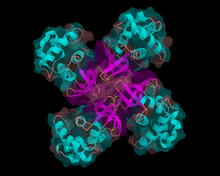
Structure of E. coli helicase RuvA
DNA helicase
Identifiers
EC number 3.6.4.12
Databases
IntEnz IntEnz view
BRENDA BRENDA entry
ExPASy NiceZyme view
KEGG KEGG entry
MetaCyc metabolic pathway
PRIAM profile
PDB structures RCSB PDB PDBe PDBsum
RNA helicase
Identifiers
EC number 3.6.4.13
Databases
IntEnz IntEnz view
BRENDA BRENDA entry
ExPASy NiceZyme view
KEGG KEGG entry
MetaCyc metabolic pathway
PRIAM profile
PDB structures RCSB PDB PDBe PDBsum
Helicases are a class of enzymes vital to all living organisms. Their main function is to unpackage an organism's genes. They are motor proteins that move directionally along a nucleic acid phosphodiester backbone, separating two annealed nucleic acid strands (i.e., DNA, RNA, or RNA-DNA hybrid) using energy derived from ATP hydrolysis. There are many helicases resulting from the great variety of processes in which strand separation must be catalyzed. Approximately 1% of eukaryotic genes code for helicases.[1] In humans, 95 non-redundant helicases are coded for in the genome, 64 RNA helicases and 31 DNA helicases.[2] Many cellular processes, such as DNA replication, transcription, translation, recombination, DNA repair, and ribosome biogenesis involve the separation of nucleic acid strands that necessitates the use of helicases.
 KEEFE ET AL" Bloom syndrome occurs most frequently in the Ashkenazi Jewish population with patients almost exclusively homozygous for a frameshift mutation resulting from a 6 bp deletion/7 bp insertion at nucleotide 2,281 (BLMAsh). This mutation causes premature termination of the encoded gene product producing a truncated protein of 739 amino acids while the full length protein contains 1417 amino acids.
The mutated gene in Bloom syndrome, BLM, was localized to chromosome 15q26.1 and encodes a member of the RecQ family of DNA helicases. This family also contains several other genes that are associated with disease phenotypes including the Werner Syndrome protein (WRN) and the defective protein in Rothmund-Thomson syndrome (RecQL4). Both of these diseases also feature an increased incidence of cancer. BLM, along with the rest of the members of this family, exhibits 3'-5' helicase activity and plays a role in DNA repair and recombination. BLM functions during replication stress and is required for the recruitment of several other important repair proteins including NBS1, BRCA1, Rad51 and MLH1. In addition, the BLM helicase is involved in recombinational repair events as evidenced by its ability to promote branch migrations of Holliday junctions at stalled replication forks. BLM may also play a role in apoptosis since it directly interacts with p53 and helps regulate its transcriptional activity."
=========================================================================

3.ATM gene
Ataxia telangiectasia mutated
From Wikipedia, the free encyclopedia
Jump to: navigation, search
Ataxia telangiectasia mutated
Identifiers
Symbols ATM; AT1; ATA; ATC; ATD; ATDC; ATE; TEL1; TELO1
External IDs OMIM607585 MGI107202 HomoloGene30952 ChEMBL: 3797 GeneCards: ATM Gene
EC number 2.7.11.1
Orthologs
Species Human Mouse
Entrez 472 11920
Ensembl ENSG00000149311 ENSMUSG00000034218
UniProt Q13315 Q62388
RefSeq (mRNA) NM_000051 NM_007499
RefSeq (protein) NP_000042 NP_031525
Location (UCSC) Chr 11:
108.09 – 108.24 Mb
Chr 9:
53.44 – 53.54 Mb

PubMed search [1] [2]
Ataxia telangiectasia mutated (ATM) is a serine/threonine protein kinase that is recruited and activated by DNA double-strand breaks. It phosphorylates several key proteins that initiate activation of the DNA damage checkpoint, leading to cell cycle arrest, DNA repair or apoptosis. Several of these targets, including p53, CHK2 and H2AX are tumor suppressors.
The protein is named for the disorder Ataxia telangiectasia caused by mutations of ATM.[1]

GOLDGAR ET AL suggested:

"The risk estimates from this study suggest that women carrying the pathogenic variant, ATM c.7271T > G, or truncating mutations demonstrate a significantly increased risk of breast cancer with a penetrance that appears similar to that conferred by germline mutations in BRCA2."

=============================================================

4. MRE 11 gene

MRE11A

From Wikipedia, the free encyclopedia
Jump to: navigation, search
MRE11 meiotic recombination 11 homolog A (S. cerevisiae)
Available structures
PDB Ortholog search: PDBe, RCSB
Identifiers
Symbols MRE11A; ATLD; HNGS1; MRE11; MRE11B
External IDs OMIM600814 MGI1100512 HomoloGene4083 GeneCards: MRE11A Gene
RNA expression pattern
PBB GE MRE11A 205395 s at tn.png
More reference expression data
Orthologs
Species Human Mouse
Entrez 4361 17535
Ensembl ENSG00000020922 ENSMUSG00000031928
UniProt P49959 Q61216
RefSeq (mRNA) NM_005590 NM_018736
RefSeq (protein) NP_005581 NP_061206
Location (UCSC) Chr 11:
94.15 – 94.23 Mb
Chr 9:
14.78 – 14.84 Mb

PubMed search [1] [2]
Double-strand break repair protein MRE11A is a protein that in humans is encoded by the MRE11A gene.[1]
This gene encodes a nuclear protein involved in homologous recombination, telomere length maintenance, and DNA double-strand break repair. By itself, the protein has 3' to 5' exonuclease activity and endonuclease activity. The protein forms a complex with the RAD50 homolog; this complex is required for nonhomologous joining of DNA ends and possesses increased single-stranded DNA endonuclease and 3' to 5' exonuclease activities. In conjunction with a DNA ligase, this protein promotes the joining of noncomplementary ends in vitro using short homologies near the ends of the DNA fragments. This gene has a pseudogene on chromosome 3. Alternative splicing of this gene results in two transcript variants encoding different isoforms.[2]

Interactions

MRE11A has been shown to interact with Ku70,[3] Ataxia telangiectasia mutated,[4][5] MDC1,[6] Rad50,[3][5][7][8][9] Nibrin,[5][9][10][11][12] TERF2[13] and BRCA1.[5][7][14][15]

 FUKUDA ET AL

"MRE11, RAD50, and XRS2 have been identified in yeast as components of the HR and NHEJ pathways (4) . A physical complex with these proteins has been identified. In vertebrates, MRE11 and RAD50 form a complex with NBS1, whose mutation causes NBS (5 , 6) . The clinical features of NBS overlap with those of AT. They are characterized by chromosome instability, increased hypersensitivity to ionizing radiation, immunodeficiency, and predisposition to cancer. AT is caused by mutations in the ATM gene, which encodes a protein kinase homologous with phosphatidylinositol-3 kinase (7) . ATM is a key regulator of the cellular response to DSBs. NBS1 is phosphorylated in an ATM-dependent manner after ionizing radiation, suggesting a link between ATM and NBS1 in a common signaling pathway (8) . MRE11 phosphorylation upon DNA damage is dependent on NBS1 (9) . Therefore, it is highly likely that MRE11 participates in the same pathway in response to DNA damage. Consistent with this functional interaction, hypomorphic mutations in the MRE11 gene cause ataxia-telangiectasia-like disorder, the phenotypes of which are indistinguishable from those of AT (10) ."

 ATM mutations play a causal role in AT and have been demonstrated in lymphoid malignancies
======================================================================

5.  RAD51
From Wikipedia, the free encyclopedia
Jump to: navigation, search
RAD51 homolog (S. cerevisiae)

A filament of Rad51 based on PDB 1SZP.[1]
Available structures
PDB Ortholog search: PDBe, RCSB
Identifiers
Symbols RAD51; BRCC5; HRAD51; HsRad51; HsT16930; MRMV2; RAD51A; RECA
External IDs OMIM179617 MGI97890 HomoloGene2155 GeneCards: RAD51 Gene
RNA expression pattern
PBB GE RAD51 205024 s at tn.png
PBB GE RAD51 205023 at tn.png
More reference expression data
Orthologs
Species Human Mouse
Entrez 5888 19361
Ensembl ENSG00000051180 ENSMUSG00000027323
UniProt Q06609 Q08297
RefSeq (mRNA) NM_001164269 NM_011234
RefSeq (protein) NP_001157741 NP_035364
Location (UCSC) Chr 15:
40.99 – 41.02 Mb
Chr 2:
119.11 – 119.15 Mb

PubMed search [1] [2]
"RAD51 is an eukaryote gene. The protein encoded by this gene is a member of the RAD51 protein family which assist in repair of DNA double strand breaks. RAD51 family members are homologous to the bacterial RecA and yeast Rad51. The protein is highly conserved in most eukaryotes, from yeast to humans.
BRCA genes

This protein can interact with the ssDNA-binding protein RPA, BRCA2, PALB2[3] and RAD52.
The structural basis for Rad51 filament formation and its functional mechanism still remain poorly understood. However, recent studies using fluorescent labeled Rad51[4] has indicated that Rad51 fragments elongate via multiple nucleation events followed by growth, with the total fragment terminating when it reaches about 2 μm in length. Disassociation of Rad51 from dsDNA, however, is slow and incomplete, suggesting that there is a separate mechanism that accomplishes this."

"The RAD51 gene family, genetic instability and cancer.

Source

Medical Research Council, Radiation and Genome Stability Unit, Harwell, Oxfordshire OX11 0RD, UK. j.thacker@har.mrc.ac.uk

Abstract

Inefficient repair or mis-repair of DNA damage can cause genetic instability, and defects in some DNA repair genes are associated with rare human cancer-prone disorders. In the last few years, homologous recombination has been found to be a key pathway in human cells for the repair of severe DNA damage such as double-strand breaks. The RAD51 family of genes, including RAD51 and the five RAD51-like genes (XRCC2, XRCC3, RAD51L1, RAD51L2, RAD51L3) are known to have crucial non-redundant roles in this pathway."---------------------------------------------------------------------

THE BRCA ITSELF TO WORK NEED A BUNCH OF VARIOUS COFACTORS CALLED  FANCB-Related Fanconi Anemia, FANCC-Related Fanconi Anemia, FANCD2-Related Fanconi Anemia, FANCE-Related Fanconi Anemia, FANCF-Related Fanconi Anemia, FANCG-Related Fanconi Anemia, FANCI-Related Fanconi Anemia, FANCL-Related Fanconi Anemia, FANCM-Related Fanconi Anemia, PALB2-Related Fanconi Anemia, RAD51C-Related Fanconi Anemia, SLX4-Related Fanconi Anemia (ALTER ET AL!)

========================================================================
IN OTHER NEWS...
HPV Vaccine: 2 Doses as Good as 3 Doses in Young Women

Troy Brown.  go to article!
IMPLICATIONS OF TARGET THERAPY ON BONE MARROW FAILURE SYNDROMES:

As we move forward, and with the increase of therapeutic agents available in our armamentarium, the focus of therapy in bone marrow failure syndrome will be on the gene altered rather then putting these disease in a global group.  Indeed a Myelodysplastic Syndrome driven by alteration of gene repair mechanisms (BRCA or RAD51) should not be treated as a Ribosomal derangement or a change in a gene that induce morphogenic changes (Dyskeratosis Congenita).  We know by now that PARP inhibitors have a better chances theoritically to act BRCA alterations then immunomodulators (Thalidomid, Revlimid) which should be attempted in Dyskeratosis like syndrome.
We know now that marrow failure syndromes particularly the congenital one have failure or gene alterations in the following targets:

1. Co-factors to DNA repair genes (Fanconi Anemia) (PARP inhibitor)
2. Telomere biology (may be the MTOR inhibitor)
3. Morphogenesis  (Revlimid, Thalidomid, Anti MEK)
4. Receptors of growth factors (Erythropoietin, thrombopoietin ) (MPL gene) (Interferon)
5. Ribosomal biogenesis (antibiotics)
6. Histone  (Acetyl transferaseinhibitor)
7. Mitochondrial Gene (MTOR Inhibitor)

Proof of concept is still needed in some cases
but that this is the way to go, we need more discernment
MDS, first obtain gene altered and make a therapeutic decision!

Tuesday, April 30, 2013

KNOW MORE ABOUT SUTENT


Indication
SUTENT® (sunitinib malate) is indicated for the treatment of advanced renal cell carcinoma (RCC).
Important Safety Information
Hepatotoxicity has been observed in clinical trials and post-marketing experience. This hepatotoxicity may be severe, and deaths have been reported. Monitor liver function tests before initiation of treatment, during each cycle of treatment, and as clinically indicated. SUTENT should be interrupted for Grade 3 or 4 drug-related hepatic adverse events and discontinued if there is no resolution. Do not restart SUTENT if patients subsequently experience severe changes in liver function tests or have other signs and symptoms of liver failure.
Women of childbearing potential should be advised of the potential hazard to the fetus and to avoid becoming pregnant.
Given the potential for serious adverse reactions (ARs) in nursing infants, a decision should be made whether to discontinue nursing or SUTENT.
Cardiovascular events, including heart failure, myocardial disorders, and cardiomyopathy, some of which were fatal, have been reported. Monitor patients for signs and symptoms of congestive heart failure (CHF) and, in the presence of clinical manifestations, discontinuation is recommended. Patients who presented with cardiac events, pulmonary embolism, or cerebrovascular events within the previous 12 months were excluded from clinical studies.
SUTENT has been shown to prolong QT interval in a dose-dependent manner, which may lead to an increased risk for ventricular arrhythmias including torsades de pointes, which has been seen in <0.1% of patients. Monitoring with on-treatment electrocardiograms and electrolytes should be considered.
Hypertension may occur. Monitor blood pressure and treat as needed with standard antihypertensive therapy. In cases of severe hypertension, temporary suspension of SUTENT is recommended until hypertension is controlled.
There have been rare (<1%) nonfatal reports of subjects presenting with seizures and radiological evidence of reversible posterior leukoencephalopathy syndrome (RPLS).
Hemorrhagic events, including tumor-related hemorrhage such as pulmonary hemorrhage, have occurred. Some of these events were fatal. Perform serial complete blood counts (CBCs) and physical examinations.
Osteonecrosis of the jaw (ONJ) has been reported. Consider preventive dentistry prior to treatment with SUTENT. If possible, avoid invasive dental procedures, particularly in patients receiving bisphosphonates.
Cases of tumor lysis syndrome (TLS) have been reported primarily in patients with high tumor burden. Monitor these patients closely and treat as clinically indicated.
Thyroid dysfunction may occur. Monitor thyroid function in patients with signs and/or symptoms of hypothyroidism or hyperthyroidism and treat per standard medical practice.
Cases of impaired wound healing have been reported. Temporary interruption of therapy with SUTENT is recommended in patients undergoing major surgical procedures.
Adrenal hemorrhage was observed in animal studies. Monitor adrenal function in case of stress such as surgery, trauma, or severe infection.
CBCs with platelet count and serum chemistries including phosphate should be performed at the beginning of each treatment cycle for patients receiving treatment with SUTENT.
Dose adjustments are recommended when administered with CYP3A4 inhibitors or inducers.
The most common ARs occurring in ≥20% of patients receiving SUTENT for treatment-naïve metastatic RCC (all grades, vs IFNα) were diarrhea (66% vs 21%), fatigue (62% vs 56%), nausea (58% vs 41%), anorexia (48% vs 42%), altered taste (47% vs 15%), mucositis/stomatitis (47% vs 5%), pain in extremity/limb discomfort (40% vs 30%), vomiting (39% vs 17%), bleeding, all sites (37% vs 10%), hypertension (34% vs 4%), dyspepsia (34% vs 4%), arthralgia (30% vs 19%), abdominal pain (30% vs 12%), rash (29% vs 11%), hand-foot syndrome (29% vs 1%), back pain (28% vs 14%), cough (27% vs 14%), asthenia (26% vs 22%), dyspnea (26% vs 20%), skin discoloration/yellow skin (25% vs 0%), peripheral edema (24% vs 5%), headache (23% vs 19%), constipation (23% vs 14%), dry skin (23% vs 7%), fever (22% vs 37%), and hair color changes (20% vs <1%). The most common grade 3/4 ARs (occurring in ≥5% of patients with RCC receiving SUTENT vs IFNα) were fatigue (15% vs 15%), hypertension (13% vs <1%), asthenia (11% vs 6%), diarrhea (10% vs <1%), hand-foot syndrome (8% vs 0%), dyspnea (6% vs 4%), nausea (6% vs 2%), back pain (5% vs 2%), pain in extremity/limb discomfort (5% vs 2%), vomiting (5% vs 1%), and abdominal pain (5% vs 1%).
The most common grade 3/4 lab abnormalities (occurring in ≥5% of patients with RCC receiving SUTENT vs IFNα) included lymphocytes (18% vs 26%), lipase (18% vs 8%), neutrophils (17% vs 9%), uric acid (14% vs 8%), platelets (9% vs 1%), hemoglobin (8% vs 5%), sodium decreased (8% vs 4%), leukocytes (8% vs 2%), glucose increased (6% vs 6%), phosphorus (6% vs 6%), and amylase (6% vs 3%).
PFIZER!
TARGET THERAPY IS ON TARGET!


Skip Navigation Links ONLINE FIRST: Phase II Mantle Cell Lymphoma Study Shows 100%...
Online First/Online Only
Articles/items published ahead of print or only online.
Wednesday, April 24, 2013
ONLINE FIRST: Phase II Mantle Cell Lymphoma Study Shows 100% Response with Epigenetic Therapy

BY ROBERT H. CARLSON

WASHINGTON -- Hematologic malignancies set the precedent for conventional therapies of cancer many years ago in terms of combination chemotherapy, and they continue to do so with some exciting new trends in therapy.

In one report here at the American Association for Cancer Research an epigenetic/immunotherapy regimen of cladribine, rituximab, and vorinostat produced a 100 percent response rate with 86 percent complete remissions in newly diagnosed mantle cell lymphoma.
======================================
YOU CANNOT BEAT 100%!
=====================================

JUST A TASTE OF THE DEBATE, GO TO ORIGINAL ARTICLE AND READ IT!

Medscape Medical News from the:

This coverage is not sanctioned by, nor a part of, the European Society of Endocrinology.

To [Vitamin] D or Not to D? That Is the Question

Lisa Nainggolan
Apr 29, 2013
 
COPENHAGEN, Denmark — Two leading experts in the field of vitamin D agreed to disagree yesterday here at the 2013 European Congress on Endocrinology during a lighthearted debate on the subject of whether or not everyone needs more vitamin D.
But their arguments were backed up by some serious science, and they both concurred that there are certain groups of people in whom it is necessary to ensure that vitamin-D levels are sufficient, such as pregnant women and those at risk for or with osteoporosis. And they also agreed on one way people can obtain more vitamin D: by going out in the sun for 30 minutes per day.
Where they differed, however, was that the vitamin-D proponent, Chantal Mathieu, MD, from Catholic University, Leuven, Belgium, said the list of people who need sufficient vitamin D "is so long that it really just makes more sense to give everyone small doses."
In the opposite corner, however, Mark Cooper, MD, from University Hospital, Birmingham, United Kingdom, argued that it is really only necessary to supplement specific, at-risk groups of people. "I am an investigator in randomized clinical trials of vitamin D, and I have nothing personally against [it], and I use it in my patients. But I tend to give it to people who actually need it, and that doesn't really include most of us," he observed.
And Dr. Cooper — who noted that there is a huge sector of the scientific community that is "evangelical" in its pro–vitamin-D stance — warned that physicians have been here before, with many other nutrients that subsequently, in large intervention trials, turned out to have a null effect or even be harmful. In fact, there is already evidence of risks with supplements of vitamin D from randomized clinical trials, with no evidence of benefit, he argued.
"Vitamin D — we all need more? Most of us don't, and more could actually do more harm than good."
What Does Vitamin D Do, and How Is "Deficiency" Defined?
Dr. Mathieu said the key role of vitamin D "is to promote resorption of calcium via the gut. One big lesson from all of the literature is that vitamin-D deficiency is not only bad because it's vitamin-D deficiency, but it also creates a bad calcemic status."
Vitamin-D deficiency is generally defined as a level of less than 20 ng/mL (<50 nm/L), and there are correlations in large observational studies "indicating that if you are vitamin-D deficient you get more cancer, especially colon cancer, you get more cardiovascular diseases, your immune system doesn't function properly, and overall you have a higher risk of dying," she stressed.
Going out in the sun is one option to boost vitamin D, she explained, noting that "even the dermatologists in Australia have reversed their zero-tolerance stance on the sun" in the past 2 years and conceded that 15 to 30 minutes per day in the sun "is allowed because it gives benefits." Nevertheless, the benefits must be balanced with the risks, she added, noting that "it's exactly the same wavelength of UV that you need to make vitamin D that also causes skin damage, aging, and skin cancers. So go back to nature and expose yourself to the sun, but do it with caution."
And she noted that UV rays in Northern Hemisphere winters are not strong enough to produce adequate levels of vitamin D, regardless of how long is spent in the sun. In addition, darker-skinned people, particularly those who do not expose themselves to the sun or who cover themselves, are particularly at risk. "We still see rickets in my country, in dark-skinned children who are exclusively breast-fed and whose mothers avoid the sun or are covered," she observed.
"You can also take it from foods," she explained, but added that the "only really rich food source" of vitamin D is cod-liver oil. "Salmon and mackerel from the ocean is a good source"; however, most of this fish is now bred in farms, and farm-bred fish do not have a lot of vitamin D.
 
If our skin cannot make enough vitamin D under the UV, just give vitamin D, give the hormone. Dr. Chantal Mathieu
 
"So what do we do? We are endocrinologists. If the thyroid fails, we give thyroid-hormone substitution. If our skin cannot make enough vitamin D under the UV, just give vitamin D, give the hormone itself."
But the key, she said, is to use smaller doses of vitamin D than have previously been recommended. The US Endocrine Society guidance, for example, advises supplementation with up to 2000 IU per day, but this is overzealous, she said. "A more reasonable dose is 600 to 800 IU per day," she noted, adding that she is an author on a new guidance, soon to be published in the Journal of Clinical Endocrinology and Metabolism, which will state that 2000 IU per day "is not warranted."

YOU TOO ARE INVITED!

Exclusive Interview - Broad Institute's Brian Haas on RNA-Seq
Inbox
x

Izzy Scott-Moncrieff
Apr 26 (4 days ago)

to me
  Dear MUTOMBO,


I’m getting in touch to share some hot off the press, exclusive RNA Sequencing content with you. 

I recently picked the impressive brains of Brian Haas (Manager of Genome Annotation, Outreach, Bioinformatics and Analysis at the Broad Institute) to learn more about the exciting prospects for RNA-Seq technology. You can download the full interview here.

With RNA-Seq 2013 just around the corner, (well, Boston, 18 - 20 June) and with Brian on the agenda and hosting an interactive workshop I thought you may like a sneak peek into what he will be sharing.
 
Brian is currently the lead developer of Trinity software at the Broad Institute - a novel method for the efficient and robust de novo reconstruction of transcriptomes from RNA-seq data. 

I learned that despite some really exciting developments since the emergence of RNA-Seq technology, big challenges still remain. One of the biggest of which is identifying what tools to use, how to use them, and what the requirements are in terms of bioinformatics skills, hardware and compute resources. He is making it the mission of his group at the Broad to find the answers to the bioinformatics challenges that seem to be restricting the genomics field.

You can download the interview for free here and read more about the really exciting research his team is focussing on. 

Brian is one of the expert speakers at RNA-Seq 2013, co-leading a workshop with Cole Trapnell and giving a presentation on the program with the likes of Mark Gerstein (Yale), Thomas Wu (Genentech) and Edward Oakeley (Novartis).

You can have a look at the full speaker line up and program on the website or if you download the brochure

We are closing early registration on Friday 10th May, so make sure you register before then to get best rates. Also workshops places are limited and filling up fast. To secure your place at the meeting for RNA-Seq pioneers visit www.rna-seqsummit.com/register  

Please don’t hesitate to get in touch with any questions about the meeting. It would be great to hear from you.

Best regards, 

Izzy

Izzy Scott-Moncrieff
Hanson Wade


Dr. Susan M. Nedza
Case Study

A 52 year old gentleman was diagnosed with stage IV pancreatic cancer with liver and lung metastases. His initial therapy included FOLFIRINOX, a 5-FU based combination with irinotecan, leucovorin and oxaliplatin. Imaging studies showed disease progression after 3 months of treatment. The oncologist is now requesting bevacizumab (Avastin), and plans to add it to the current regimen.

Background
Pancreatic cancer is a highly lethal malignancy, with a 5 year overall survival rate of 5%. In the setting of metastatic disease, this cancer is uniformly fatal, with a median survival of approximately 6 months. Treatment has been used primarily as a palliative measure, for clinical benefit and symptom improvement.

Gemcitabine combinations
Gemcitabine has been the backbone of pancreatic cancer treatment since 1996, and multiple combinations have been studied since that time. Unfortunately, very few treatments have led to a significant improvement in overall survival. A recent exception has been the use of gemcitabine in combination with nab-paclitaxel (Abraxane® ). The MPACT trial, which compared this combination with single agent gemcitabine, showed an increase in overall survival with combination therapy (8.5 months vs. 6.7 months).

FOLFIRINOX
A randomized, phase III trial (PRODIGE) published in 2011 evaluated the combination (FOLFIRINOX) versus single agent gemcitabine in patients with metastatic pancreatic cancer and good performance status. Combination therapy demonstrated significant improvement in progression-free survival and median overall survival, but with significantly greater toxicity than gemcitabine alone. This regimen has been added to the National Comprehensive Cancer Network (NCCN) guidelines as a category 1 recommendation for first-line treatment of metastatic pancreatic cancer in patients with good functional status.

Bevacizumab (Avastin®)
The introduction of bevacizumab (Avastin®) has been groundbreaking in cancer treatment, demonstrating efficacy in colorectal cancer and lung cancer, among others. Side effects are considered to be generally mild, with high blood pressure and mild nosebleeds among the most common. Though rare, Avastin® has also been linked to gastrointestinal perforation, thrombosis, and fatal hemorrhage.

With respect to pancreatic cancer, however, randomized phase III studies of gemcitabine in combination with Avastin® have failed to demonstrate improvements in overall survival, progression-free survival, or overall response rates. Furthermore, there was a significant increase in hypertension and proteinuria, though no toxic deaths were reported.

Discussion

In our case, the patient has received a 5-FU-based regimen with disease progression. To date, there have been no published studies demonstrating the efficacy or safety of adding Avastin® to this regimen. Because of its efficacy in other tumor types and some activity demonstrated in Phase I/II trials, this drug is often requested off-label.

Current consensus opinion would recommend either gemcitabine/gemcitabine combination or a 5FU-based regimen for first-line treatment. Upon progression, NCCN recommends a change to a regimen with a different backbone. In the case presented, changing to a gemcitabine-based regimen would be the recommended approach. Current evidence does not support the use of bevacizumab (Avastin®) for pancreatic cancer.

In cases such as this, a pathways program can guide the ordering physician in selecting an evidence-supported approach and discourage continuation of a failing regimen. We believe that providing physicians with advanced cancer treatment decision support will translate into improved, cost-effective care.

Learn more about AIM’s Integrated Oncology Solution here.

This email was sent by: AIM Specialty Health
8600 W. Bryn Mawr Ave. Ste. 800 Chicago, IL, 60631, USA
     

TRIPLE NEGATIVE BREAST CANCER TREATMENT

The reading about mechanisms of resistance of to Taxol calls for a new strategy for treatment of triple negative breast cancer; while it is true that PARP inhibitor should still be considered in BRCA positive cancers, adding AURORA inhibitors seems to offer logically the best opportunity to increase the activity of proposed first line drugs.

Indeed, triple negative breast cancer assumes that the receptors to conventional stimulants of the breast cancer cell are not functional or responsive.  Therefore, increasing the role of a direct attack of either the nuclear material or the microfilament/microtubule.  Taxol - Cisplatin combination achieves that!  Adding Avastin and other receptor stimulators could be a riskier proposition if you assume a questionable sensitivity of receptor in general.  Your best bet is an action on the Histones and further DNA destruction.  The cell division is your focus here and this is re-emphasized by the importance of CDKs as described by MD Anderson researchers.  As a matter of fact, the AURORA inhibitors by binding to Adenine and to the Histone appear to offer a potential and logical choice to recruit in first line to boost response rates!  So, pending proof of concept, we support the idea of adding Aurora inhibitors to a Taxane-Cisplatin core combination. Some of the Aurora Kinase also target CDKs and JAK2.  These will be my choice for new trials!

After the cancer has seen chemotherapy, endothelial cells have been altered, hypoxia has been triggered by closure of some of the blood vessel closure, the MTOR has been stimulated, we believe adding the MTOR makes more sense.  This has been also suggested after failure of Avastin,  These concepts have been publicized, It is time to move to clinical trial! (FOR THOSE WHO CAN, WE HAVE OUR HANDS TIED BY HUMAN HISTORY!)