Friday, March 22, 2013

Meat consumption and mortality - results from the European Prospective Investigation into Cancer and Nutrition


Results
As of June 2009, 26,344 deaths were observed. After multivariate adjustment, a high consumption of red meat was related to higher all-cause mortality (hazard ratio (HR) = 1.14, 95% confidence interval (CI) 1.01 to 1.28, 160+ versus 10 to 19.9 g/day), and the association was stronger for processed meat (HR = 1.44, 95% CI 1.24 to 1.66, 160+ versus 10 to 19.9 g/day). After correction for measurement error, higher all-cause mortality remained significant only for processed meat (HR = 1.18, 95% CI 1.11 to 1.25, per 50 g/d). We estimated that 3.3% (95% CI 1.5% to 5.0%) of deaths could be prevented if all participants had a processed meat consumption of less than 20 g/day. Significant associations with processed meat intake were observed for cardiovascular diseases, cancer, and 'other causes of death'. The consumption of poultry was not related to all-cause mortality.
Conclusions
The results of our analysis support a moderate positive association between processed meat consumption and mortality, in particular due to cardiovascular diseases, but also to cancer.
Keywords:

Socioeconomic patient characteristics predict delay in cancer diagnosis: a Danish cohort study


http://www.biomedcentral.com/1472-6963/8/49

JAMA Network | JAMA | DELAY IN DIAGNOSIS AND TREATMENT OF CANCER

This is an article from 1950 that already pointed out what matters to improve patient's survival outcomes:

JAMA Network | JAMA | DELAY IN DIAGNOSIS AND TREATMENT OF CANCER

Michaelson Adv Imm Onc 2 12 07 pdf free ebook download from www.lifemath.net

Michaelson Adv Imm Onc 2 12 07 pdf free ebook download from www.lifemath.net

Thursday, March 21, 2013

The abyss between cancer diagnosis and start of treatment

The outcome for the newly diagnosed patient very much depends on how high the awareness of the diagnosing physician is for his type of cancer, how deep and fresh his knowledge is about current treatment guidelines and options, and on how strong a network of specialized oncologists he can rely on to refer his patient to. If one of these links is weak, the trials and tribulations are only just beginning to start.
Right from from Day 1 should Cancer be an integral part of the Medical School Curriculum  and not pushed down the list of priorities and for specialization only many years later.
Cancer comes in all shapes and sizes and can occur at all ages. Being able to recognize it at its earliest stage will dramatically improve patient survival and the patient's quality of life, and minimize the premature loss of human lives immensely.

IXL Gene in pancreatic cancer

IXL, Intersex like cell, a survival regulator, located on 19q13,
note the q location of the amplicon ( a piece of DNA or RNA that is the source and/or product of natural or artificial amplification or replication events) the 10% rate of amplification is clearly higher than the usual few, with 5% of secondary amplification making this a clear Amplicon.  Knocking down this stops cells in Go-G1 per Kuuselo et al.  As the Component of the Mediator complex, it is a co-activator that  regulates the transcription of nearly all RNA polymerase II-dependent genes which are at the origin of m-RNA formation.  It puts this gene at the initiation complex.  At the sole of enzyme fabrication, regulator fabrication, and formation of transcription factors.  Tinkering with gene blocks transcription. That's it!  Even splicing will in fact be affected to some extent.

This gene interacts with AP-1

and

AP-1 transcription factor

From Wikipedia, the free encyclopedia
Jump to: navigation, search

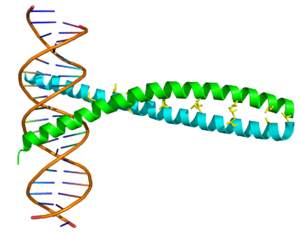
AP-1/DNA complex. Crystallographic structure (PDB 1FOS) of the AP-1 heterodimer comprising c-Fos (cyan) and c-Jun (green) complexed with DNA (brown).
In the field of molecular biology, the activator protein 1 (AP-1) is a transcription factor which is a heterodimeric protein composed of proteins belonging to the c-Fos, c-Jun, ATF and JDP families. It regulates gene expression in response to a variety of stimuli, including cytokines, growth factors, stress, and bacterial and viral infections.[1] AP-1 in turn controls a number of cellular processes including differentiation, proliferation, and apoptosis.[2]
AP-1 upregulates transcription of genes containing the TPA DNA response element (TRE; 5'-TGAG/CTCA-3').[1] AP-1 binds to this DNA sequence via a basic amino acid region, while the dimeric structure is formed by a leucine zipper.[3] 

SO HERE PANCREATITIS, ALCOHOL, VIRUSES AND OTHER STRESSORS FIND THEIR WAY TO THE PATHOGENESIS OF PANCREATIC CANCERS!

It is also connected to

CUTL1

and it needs CUTL-1 now to have muscle, but unleash the powerful regulator it encloses.  It may use this regulator to silence other genes that may lead to apoptosis. The IXL gene is a strategist in its advancement of pancreatic cancer.  This is a major target!

And 3rd, and not the least 

it interferes with ATF2

'This gene encodes a transcription factor that is a member of the leucine zipper family of DNA-binding proteins. This protein binds to the cAMP-responsive element (CRE), an octameric palindrome. The protein forms a homodimer or heterodimer with c-Jun and stimulates CRE-dependent transcription. The protein is also a histone acetyltransferase (HAT) that specifically acetylates histones H2B and H4 in vitro; thus, it may represent a class of sequence-specific factors that activate transcription by direct effects on chromatin components. Additional transcript variants have been identified but their biological validity has not been determined.[1]
The gene atf2 is located at human chromosome 2q32.[2] '  (wikipedia

BELIEVE ME WHEN THEY SAY LEUCINE, THE MTORs ARE NOT FAR BEHIND!!!

BETA 4 INTEGRIN: a gene that does more than being an adhesion molecules it is the road to a poorly described and not well recognized pathway

The LysRS-Ap4A-MITF signaling pathway
The LysRS-Ap4A-MITF signaling pathway was first discovered in mast cells, in which , the MAPK pathway is activated upon allergen stimulation. Lysyl-tRNA synthetase (LysRS), which normally resides in the multisynthetase complex with other tRNA sythetases, is phosphorylated on Serine 207 in a MAPK-dependent manner.[30] This phosphorylation causes LysRS to change its conformation, detach from the complex and translocate into the nucleus, where it associates with the MITF-HINT1 inhibitory complex. The conformational change switches LysRS activity from aminoacylation of Lysine tRNA to diadenosine tetraphosphate (Ap4A) production. Ap4A binds to HINT1, which releases MITF from the inhibitory complex, allowing it to transcribe its target genes.[31] Activation of the LysRS-Ap4A-MITF signaling pathway by isoproterenol has been confirmed in cardiomyocytes, where MITF is a major regulator of cardiac growth and hypertrophy.[32][33](wikipedia)

Not only it gives Hypertrophy but epidermolysis goes through this intergrin, it participates in the ERBB pathways.  Mark my word this is are critical pathways in pancreatic cancers.

MTIF GIVES YOU MOTIVES TO GO AFTER IT!
MAKING THE ERBIN A PLAUSIBLE TARGET.
MAKING ALSO A STRONGER CASE THAT MEMBRANE CYTOSKELETON SHOULD BE A GOOD TARGET BECAUSE OF THE WAY IT DRIVES ITS PATHWAY NOT THROUGH THE CYTOSOL( ALTHOUGH THERE IS A SECONDARY RAS/MAPK STIMULATION,) BUT THE PATHWAY HERE IS THROUGH THE RETICULUM ENDOTHELIUM DIRECTLY TO THE NUCLEUS!  CONCEPTUALLY, AN ANTIBODY TO LAMININ ATTACHED TO A SUBUNIT OF A LIPOLYTIC COMPOUND SHOULD HAVE A THERAPEUTIC OR CHEMICAL EFFECT AT THIS LEVEL.  AN INTERESTING APPROACH.  CHANCES ARE IT MAY ALSO HAVE A STRONG IMPACT ON THE WNT-PATHWAY WHICH TRAVEL CLOSE BY AND IS IMPORTANT IN BREAST CANCER!

MTA-1: THIS IS A REAL OPPORTUNITY
Here the cell stopped fooling around trying to lie to you.  Here the cell says to you this is one of my way to metastatasize.  yes this is my gene to mestastasize and I will work like any CBF like molecule by attaching to DNA and make me protein that will have me spread like wild fire!   And by the way I will use a growth hormone like Estrogen.   no kidding around
 "MTA1 has been shown to interact with HDAC1,[4][5] Histone deacetylase 2,[4][6][5] MTA2,[4] Estrogen receptor alpha[7][5] and MNAT1.[8] MTA1 has also been shown to inhibit SMAD7 at the transcriptional level[9]"  

IT DOES NEED TGF TO WORK, TGF IS FOR LOCAL GROWTH ANYWAY THAT WHY IT BLOCKS THE SMAD.

SPINT2
Mutation at SPINT2 leads to significant Malignant Ascites and peritoneal invasion, SPINT 2 is a suppressor of this phenomena. On the Intestinal membrane deficiency of SPINT2 leads to sodium induced/containing diarrhea.  This is also true in Ovarian cancer or peritoneal based tumors.  Targeting this is better then trying Avastin, a blind approach when it comes to effusions management.

MMP11

A metalloproteiase, aimed at breaking down extracellular matrix and be on the move.  Targeting MMP for cancer has proven futile.  The cell is not stupid, it does not put out things that is going to hunt it!  It first builds a strong inhibitor to metalloproteinases.  In fact lack of inhibitors has been recognized as the main pathogenesis of TTP.   With the ADAMs being the integrins involved!  and next is that Inhibitor which is of course expressed in pancreatic cancer.

TIMP1

TIMP1

From Wikipedia, the free encyclopedia
Jump to: navigation, search
TIMP metallopeptidase inhibitor 1

PDB rendering based on 1d2b.
Available structures
PDB Ortholog search: PDBe, RCSB
Identifiers
Symbols TIMP1; CLGI; EPA; EPO; HCI; TIMP
External IDs OMIM305370 MGI98752 HomoloGene36321 GeneCards: TIMP1 Gene
RNA expression pattern
PBB GE TIMP1 201666 at tn.png
More reference expression data
Orthologs
Species Human Mouse
Entrez 7076 21857
Ensembl ENSG00000102265 ENSMUSG00000001131
UniProt P01033 P12032
RefSeq (mRNA) NM_003254 NM_001044384
RefSeq (protein) NP_003245 NP_001037849
Location (UCSC) Chr X:
47.44 – 47.45 Mb
Chr X:
20.87 – 20.87 Mb

PubMed search [1] [2]
TIMP metallopeptidase inhibitor 1, also known as TIMP1, a tissue inhibitor of metalloproteinases, is a glycoprotein that is expressed from the several tissues of organisms.
This protein a member of the TIMP family. The glycoprotein is a natural inhibitor of the matrix metalloproteinases (MMPs), a group of peptidases involved in degradation of the extracellular matrix. In addition to its inhibitory role against most of the known MMPs, the encoded protein is able to promote cell proliferation in a wide range of cell types, and may also have an anti-apoptotic function.
==============
PRKCA  see PRKCG
Here Phorbol esters, diacylglycerol, and calcium become important for the cell performance of various functions.  Did I mention few targets, I truly believe I did!

CDH1  The Cadherin by excellence, not only important as adhesion molecule and role in metastasis.  Its role is amplified by what else anchors here such as Vinculin, and others molecules such as Plakoglobins, amplifying the role.  Remember even Cytochrome C is anchored at the mitochondrial membrane and its release leads to apoptosis!
The anchors are legitimate targets therefore, and brings to mind NACA1 in the anchoring to Histone deacetyl transferase (SEE OUR LEUKEMIA SECTION)  CDH13 THAT'S ANOTHER BALL GAME ALL TOGETHER.  THE CELL TWEACKS SOMETHING AND IT IS ANOTHER BALL GAME ALL TOGETHER!
==========================

Wednesday, March 20, 2013

Pancreatic cancers

Annual Incidence 43,000 new cases a year in the United States.
Annual Mortality 35, 000, making one of the deadliest cancer in the United States.
There are suggestions that Tobacco may play a role in the Occurrence.
BRCA1 and BRCA 2 have been implicated in familial cases.  Other Hereditary cases involve the HNPCC genes, p16, Ataxia Telangiectasia and Peutz-Jeghers syndrome)
KRAS, IGFR-1,DCP4, p16, p53 and BRCA2 have been implicated.
No screening method has been recommended.
The disease is clearly unresectable when Mesenteric vessels are involved or when evident metastasis are seen.

GENES IN CHORIOCARCINOMA

The major interest here at the CRBCM is of course obtaining a cure.  There is therefore a strong interest in knowing more about cancers that are deemed curable today.  Studying the genes known in these diseases could provide some clues to their susceptibility to chemotherapy drugs available today. Below are some of the genes for CHORIOCARCINOMA:

1. NECC1 on 4q11  (notice the "q" as this may be bad news)

This is a gene of differentiation.  And from what we have gathered, it is a gene that codes for a Core Binding Factor like Molecule, these complexes of major proteins with various functions put together to direct cellular functions in directions.  The proteins globally function as regulators of other cellular functions. These proteins are sequentially positioned in the CBF to drive into some directions.  Most of the times, the CBF has a portion that attaches to the DNA.  The attachment could silence the DNA.  The silenced DNA here seems to block Cardiac differentiation and forces the direction of activity toward syncytial trophoblastic differentiation.  This is why this gene is expressed in normal placenta.  In choricarcinoma, dedifferentiation occurs and NECC1 is mutated and silenced.  This is called a suppressor gene as it relates to Cardiac muscle differentiation, but clearly not for the tumor from what we gathered.  The Mutation will have to occur at the "q" location.  We will look further to establish if the "p" of this gene is located on chromosome 7.
Desactivation of NECC1 leads to cardiac hypertrophy.  Being a CBF like molecule, it may solicit Histone deacetyl transferases as part of its nuclear activity.  We still are unclear whether it is at the center of the pathogenesis of choriocarcinoma or not!
------------------------------------------------------------------------------------------------------
2.CSH1
The protein encoded by this gene is a member of the somatotropin/prolactin family of hormones and plays an important role in growth control. The gene is located at the growth hormone locus on chromosome 17 along with four other related genes in the same transcriptional orientation; an arrangement which is thought to have evolved by a series of gene duplications. Although the five genes share a remarkably high degree of sequence identity, they are expressed selectively in different tissues. Alternative splicing generates additional isoforms of each of the five growth hormones, leading to further diversity and potential for specialization. This particular family member is expressed mainly in the placenta and utilizes multiple transcription initiation sites. Expression of the identical mature proteins for chorionic somatomammotropin hormones 1 and 2 is upregulated during development, although the ratio of 1 to 2 increases by term. Mutations in this gene result in placental lactogen deficiency and Silver-Russell syndrome. [provided by RefSeq, Jul 2008]"
" ===========================================================================

3. IGFR1

"Insulin-like growth factor 1 (IGF-1), also called somatomedin C, is a protein that in humans is encoded by the IGF1 gene.[1][2] IGF-1 has also been referred to as a "sulfation factor"[3] and its effects were termed "nonsuppressible insulin-like activity" (NSILA) in the 1970s.
IGF-1 is a hormone similar in molecular structure to insulin. It plays an important role in childhood growth and continues to have anabolic effects in adults.
 IGF-1 is produced throughout life. The highest rates of IGF-1 production occur during the pubertal growth spurt. The lowest levels occur in infancy and old age.
Other IGFBPs are inhibitory. For example, both IGFBP-2 and IGFBP-5 bind IGF-1 at a higher affinity than it binds its receptor. Therefore, increases in serum levels of these two IGFBPs result in a decrease in IGF-1 activity."(Wikipedia)

IGFR1  amplification is used in autocrine faction to drive cancer growth, it acts here a as a TGF.
======================================================================= 

4. CHFR
E3 ubiquitin-protein ligase CHFR is an enzyme that in humans is encoded by the CHFR gene.[1][2][3]
(wikipedia)

"One protein that has been suggested to be part of the antephase checkpoint is Chfr (checkpoint protein with an FHA domain and ring finger; Scolnick and Halazonetis, 2000), a ubiquitin ligase that is down-regulated in several cell lines through methylation of its promoter (Mizuno et al., 2002). Chfr was originally reported to delay progress to prometaphase in the presence of colcemid (Scolnick and Halazonetis, 2000), and cells were surprisingly described as delaying with high cyclin B1-Cdk1 activity (Scolnick and Halazonetis, 2000), which conflicted with a role as part of the antephase checkpoint because cyclin B1-Cdk1 is fully activated only in late prophase. However, in Xenopus laevis extracts, Chfr is able to delay the activation of cyclin B-Cdk1, apparently by targeting the Polo-like kinase, Plx, for degradation by the proteasome (Kang et al., 2002), thereby preventing the activation of the Cdc25 phosphatase that activates Cdk1. Chfr has also been reported to affect Polo-like kinase levels in human cells in response to DNA damage (Shtivelman, 2003). But whether Chfr does target Polo for degradation or not is debatable because Chfr has been shown to conjugate ubiquitin via its lysine 63 residue (Bothos et al., 2003) that normally acts in signal transduction, especially for stress signals (Deng et al., 2000; Ulrich and Jentsch, 2000; Hofmann and Pickart, 2001; Pickart, 2001; Wang et al., 2001), rather than to target proteins to the proteasome."
Matsusaka and pines suggested.

CERTAINLY DOWN REGULATION HERE OPEN THE DOOR TO USE OF VELCADE OR THE PROTEASOME INHIBITORS.


--------------------------------------------------------------------------------------------------------
5. MUC3A"Associations of distinct variants of the intestinal mucin gene MUC3A with ulcerative colitis and Crohn's disease [1]." (WIKI) Its gene is located at 7q22  (notice the q) it has a prognosis value
But we also know that MUC1 will be more important in this cancer and shield by its mucinous product cell from cancer watching cells.

James Gum et al "Mucinous cancers are generally more extensive at diagnosis. Membrane mucins are an important class of glycoproteins with diverse structures and functions. These molecules contain extracellular domains that serve as a scaffold for O-glycosylation. The O-glycans associated with membrane mucins are thought to function in cytoprotection and have been demonstrated to confer anti-adhesion properties upon cells (1). This latter characteristic may play a role in the dissemination and spread of cancer cells. In addition to conferring these electrostatic/physical properties upon cells, membrane mucins can anchor carbohydrate moieties with specific functions. Selectin ligands associated with membrane mucin glycans, for example, play a role in cancer cell extravasation during metastases (2). Certain membrane mucins function in signal transduction as well (35). Several membrane mucins also serve as clinically important tumor antigens (6, 7)."


--------------------------------------------------------------------------------------------------------------
6. TAF7

TAF7

From Wikipedia, the free encyclopedia
Jump to: navigation, search
TAF7 RNA polymerase II, TATA box binding protein (TBP)-associated factor, 55kDa
Identifiers
Symbols TAF7; TAF2F; TAFII55
External IDs OMIM600573 MGI1346348 HomoloGene11768 GeneCards: TAF7 Gene
RNA expression pattern
PBB GE TAF7 201023 at tn.png
More reference expression data
Orthologs
Species Human Mouse
Entrez 6879 24074
Ensembl ENSG00000178913 ENSMUSG00000051316
UniProt Q15545 Q9R1C0
RefSeq (mRNA) NM_005642 NM_175770
RefSeq (protein) NP_005633 NP_786964
Location (UCSC) Chr 5:
140.7 – 140.7 Mb
Chr 18:
37.64 – 37.64 Mb

PubMed search [1] [2]
Transcription initiation factor TFIID subunit 7 also known as TAFII55 is a protein that in humans is encoded by the TAF7 gene.[1]
The intronless gene for this transcription coactivator is located between the protocadherin beta and gamma gene clusters on chromosome 5. The protein encoded by this gene is a component of the TFIID protein complex, a complex which binds to the TATA box in class II promoters and recruits RNA polymerase II and other factors. This particular subunit interacts with the largest TFIID subunit, as well as multiple transcription activators. The protein is required for transcription by promoters targeted by RNA polymerase II.[2]
TAFII55_N
Identifiers
Symbol TAFII55_N
Pfam PF04658
InterPro IPR006751
The general transcription factor, TFIID, consists of the TATA-binding protein (TBP) associated with a series of TBP-associated factors (TAFs) that together participate in the assembly of the transcription preinitiation complex. TAFII55 binds to TAFII250 and inhibits its acetyltransferase activity. The exact role of TAFII55 is currently unknown but studies have shown that it interacts with the C-jun pathway.[3] The conserved region is situated towards the N-terminal of the protein.[4] This entry talks about the N-terminal domain.
TAF7 interacts with TATA which


The TATA-binding protein (TBP) is a general transcription factor that binds specifically to a DNA sequence called the TATA box. This DNA sequence is found about 30 base pairs upstream of the transcription start site in some eukaryotic gene promoters.[1] TBP, along with a variety of TBP-associated factors, make up the TFIID, a general transcription factor that in turn makes up part of the RNA polymerase II preinitiation complex.[2] As one of the few proteins in the preinitiation complex that binds DNA in a sequence-specific manner, it helps position RNA polymerase II over the transcription start site of the gene. However, it is estimated that only 10-20% of human promoters have TATA boxes. Therefore, TBP is probably not the only protein involved in positioning RNA polymerase II.
TBP is involved in DNA melting (double strand separation) by bending the DNA by 80° (the AT-rich sequence to which it binds facilitates easy melting). The TBP is an unusual protein in that it binds the minor groove using a β sheet.
Another distinctive feature of TBP is a long string of glutamines in the N-terminus of the protein. This region modulates the DNA binding activity of the C-terminus, and modulation of DNA-binding affects the rate of transcription complex formation and initiation of transcription. Mutations that expand the number of CAG repeats encoding this polyglutamine tract, and thus increase the length of the polyglutamine string, are associated with spinocerebellar ataxia 17, a neurodegenerative disorder classified as a polyglutamine disease.[3]

BASICALLY INITIATE OR PARTICIPATE IN THE INITIATION OF OF TRANSCRIPTION/TRANSLATION
--------------------------------------------------------------------------------------------------------------
8. CDC123
9. PSMD
10. HAS2
11. CD44  a member of the EZRIN/S100P/ Villin -Complex
------------------------------------------------------------------------------------------------------
12. S100P 

S100P

From Wikipedia, the free encyclopedia
Jump to: navigation, search
S100 calcium binding protein P

PDB rendering based on 1j55.
Available structures
PDB Ortholog search: PDBe, RCSB
Identifiers
Symbols S100P; MIG9
External IDs OMIM600614 HomoloGene81743 GeneCards: S100P Gene
RNA expression pattern
PBB GE S100P 204351 at tn.png
More reference expression data
Orthologs
Species Human Mouse
Entrez 6286 n/a
Ensembl ENSG00000163993 n/a
UniProt P25815 n/a
RefSeq (mRNA) NM_005980 n/a
RefSeq (protein) NP_005971 n/a
Location (UCSC) Chr 4:
6.69 – 6.7 Mb
n/a
PubMed search [1] n/a
Protein S100-P is a protein that in humans is encoded by the S100P gene.[1][2][3]
The protein encoded by this gene is a member of the S100 family of proteins containing 2 EF-hand calcium-binding motifs. S100 proteins are localized in the cytoplasm and/or nucleus of a wide range of cells, and involved in the regulation of a number of cellular processes such as cell cycle progression and differentiation. S100 genes include at least 13 members which are located as a cluster on chromosome 1q21; however, this gene is located at 4p16. This protein, in addition to binding Ca2+, also binds Zn2+ and Mg2+. This protein may play a role in the etiology of prostate cancer.[3]

Interactions

S100P has been shown to interact with EZR.[4] which this protein serves as an intermediate between the plasma membrane and the actin cytoskeleton. It plays a key role in cell surface structure adhesion, migration, and organization.[2]

Interactions

VIL2 has been shown to interact with Sodium-hydrogen exchange regulatory cofactor 2,[3][4] Merlin,[5] SDC2,[6] CD43,[7] Fas ligand,[8][9] VCAM-1,[10] S100P,[11] ICAM3,[12][13] ICAM-1,[12] Sodium-hydrogen antiporter 3 regulator 1,[14][15] ICAM2,[12] Moesin,[8][16][17] PALLD[18] and PIK3R1.[19]

Given the multitude of interactions the EZERIN appears a critical molecules for signal propagation intra and extra-cellularly.  EZRIN the Villin (Vilain) comes into everything.  In the endothelium,  This complex of molecules is " involved in the generation and maintenance of the anchoring structure. These results provide the first characterization of an endothelial docking structure that plays a key role in the firm adhesion of leukocytes to the endothelium during inflammation." 
This is a basis for novel anti-inflammatory strategy.
It also interact with the so called Peripheral proteins of which  "some are water-soluble proteins and associate with lipid bilayers irreversibly and can form transmembrane alpha-helical or beta-barrel channels. Such transformations occur in pore forming toxins such as colicin A, alpha-hemolysin, and others. They may also occur in BcL-2 like protein , in some amphiphilic antimicrobial peptides , and in certain annexins . These proteins are usually described as peripheral as one of their conformational states is water-soluble or only loosely associated with a membrane.[11]"

----------------------------------------------------------------------------------------------------

13. delta-like1
14. STOX1
15. JAK/STAT1
16. GCM1
Chorion-specific transcription factor GCMa is a protein that in humans is encoded by the GCM1 gene.[1][2]
This gene encodes a DNA-binding protein with a gcm-motif (glial cell missing motif). The encoded protein is a homolog of the Drosophila glial cells missing gene (gcm). This protein binds to the GCM-motif (A/G)CCCGCAT, a novel sequence among known targets of DNA-binding proteins. The N-terminal DNA-binding domain confers the unique DNA-binding activity of this protein.[2]WIKIPEDIA


Chou et al "the activity of GCMa can be post-translationally regulated by protein phosphorylation, ubiquitination, and acetylation, it is unknown whether GCMa activity can be regulated by sumoylation. In this report, we investigated the role of sumoylation in the regulation of GCMa activity. We demonstrated that Ubc9, the E2 component of the sumoylation machinery...Our study demonstrates that GCMa is a new sumoylation substrate and its activity is down-regulated by sumoylation."

"


-----------------------------------------------------------------------------------------------------------
18. Cx31: Connexin, important in cell adhesion and embryogenesis.  In adult Mutation at Cx31 lead to sensory neural deafness as it contribute to synaptic integrity and Monocyte.  A legitimate secondary target in blood disease.
-----------------------------------------------------------------------------------------
also we will include
19. Endoglin
20. Syncitin
21. HCG
22. Connexin 31
23. E-Cadherin

Lets go to work!

Another thing to look at is the propensity in these diseases to develop thrombosis on BEP treatment!
Tuesday, March 19, 2013

Ovarian Cancer

*Researchers who conducted the study who conducted the use of Selumetinib in low grade serous Ovarian cancer were still puzzled because its activity did not follow presence of KRAS or BRAF. They have been wondering if it used another pathway.  But remember MEK is the revolving door to de-differentiation and to the reversal of mesengialization and as such increase tor susceptibility not only to chemotherapy drugs, but also to the secondary angiogenic potentiation of MEK.  That is, with anti-MEK, there is a down regulation of MAPK (as suggested) and therefore  the C-JUN and TGF and cyclins, but also down regulation of the VEGF!

They say " Our results suggest that selumetinib is an active agent, but not necessarily because of BRAF or KRAS mutational activation per se,” the authors concluded.
In an interview, Gershenson said that one reason for the lack of correlation could be biomarker instability. Among the 52 patients, specimens were available for only 40, mutational analysis was done in 34, and in 28 of those the tissue was from the primary therapy and not from the recurrent tumor. “The question arises, are these biomarkers stable over time or do they change, so that what you find in the primary tumor may not be what you find in the recurrent tumor,” he said."   They suggesting here that the lack of correlation could be due to a changing nature of Biomarker.  But the existence of other factors and pathways could not be be excluded!

Gene-Expression Profiling May Help Select Best Drugs for Pancreatic Cancer

share
Caroline Helwick 
---------------------------------------------------------------------------------------------------
In this study patient with pancreatic cancer had reportedly circulating cell and their genes could predict response to therapy but unfortunately they did not spell out which genes were reviewed.  We will investigate further this article...