Given our current understanding of the way they are suppressed by normal activity of P53, the aurora Kinase inhibitors should be used in cancers where P53 is clearly dys-regulated, and probably in disease with positive Prostate Stem Cell Antigen. This open the door to Sarcomas (chondrosarcoma being the most cited) and Pancreatic cancers (as well as Bladder cancers).
GADD45, a P53 dependent protein that inhibits Cdc2/Cyclin B1 could therefore be the best predictor of Aurora activity.
Quantification of CDCA8/Borealin, BIRC5/survivin, and INCENP may provide additional information on AURORA KINASE ACTIVITY. Like the core Binding Factor, these 3 Molecules form the Chromosomal Passenger Complex (CPC) which interact with POGZ, EVI5, and JTB.
Niehrs at al further define the role of GADD45 as "Gadd45 recruits nucleotide and/or base excision repair factors to gene-specific loci and acts as an adapter between repair factors and chromatin, thereby creating a nexus between epigenetics and DNA repair." Therefore explaining how P53 induced arrest is followed by DNA repair through recruitment of GADD45. When the cell is trying to repair itself with increase in GADD45, of course it wont want to die, therefore over-expression of GADD45 decrease the c-JUN, protecting therefore from TNF induced apoptosis. GADD45 is not good theoretically when you use Cisplatin or radiation for that matter!
*POGZ explains Nuclear transposition of P53 effects as it impacts SP1, a transcription factor with inteaction of all major playors in the cell including E2F1, POU2F1. YOU TARGET SP1 WITH WITH AFERIN FOR EXAMPLE, IT IS IMPOSSIBLE TO COME UP EMPTY HANDED.
*EVI5
"Identification of Rab11 as a small GTPase binding protein for the Evi5 oncogene"
This article has been cited by other articles in PMC.
Abstract
The Evi5 oncogene has recently been shown to regulate the stability and accumulation of critical G1
cell cycle factors including Emi1, an inhibitor of the
anaphase-promoting complex/cyclosome, and cyclin A. Sequence analysis of
the amino terminus of Evi5 reveals a Tre-2, Bub2, Cdc16 domain, which
has been shown to be a binding partner and GTPase-activating protein
domain for the Rab family of small Ras-like GTPases. Here we describe
the identification of Evi5 as a candidate binding protein for Rab11, a
GTPase that regulates intracellular transport and has specific roles in
endosome recycling and cytokinesis. By yeast two-hybrid analysis,
immunoprecipitation, and Biacore analysis, we demonstrate that Evi5
binds Rab11a and Rab11b in a GTP-dependent manner. However, Evi5
displays no activation of Rab11 GTPase activity in vitro. Evi5 colocalizes with Rab11 in vivo,
and overexpression of Rab11 perturbs the localization of Evi5,
redistributing it into Rab11-positive recycling endosomes.
Interestingly, in vitro binding studies show that Rab11
effector proteins including FIP3 compete with Evi5 for binding to Rab11,
suggesting a partitioning between Rab11–Evi5 and Rab11 effector
complexes. Indeed, ablation of Evi5 by RNA interference causes a
mislocalization of FIP3 at the abscission site during cytokinesis. These
data demonstrate that Evi5 is a Rab11 binding protein and that Evi5 may
cooperate with Rab11 to coordinate vesicular trafficking, cytokinesis,
and cell cycle control independent of GTPase-activating protein
function.
Keywords: cytokinesis, GTPase-activating protein, recycling endosome".
*JTB
JTB (gene)
From Wikipedia, the free encyclopedia
| Jumping translocation breakpoint | |||||||||||
|---|---|---|---|---|---|---|---|---|---|---|---|
|
|||||||||||
| Identifiers | |||||||||||
| Symbols | JTB; HJTB; HSPC222; PAR; hJT | ||||||||||
| External IDs | OMIM: 604671 MGI: 1346082 HomoloGene: 4870 GeneCards: JTB Gene | ||||||||||
|
|||||||||||
| RNA expression pattern | |||||||||||
 |
|||||||||||
 |
|||||||||||
| More reference expression data | |||||||||||
| Orthologs | |||||||||||
| Species | Human | Mouse | |||||||||
| Entrez | 10899 | 23922 | |||||||||
| Ensembl | ENSG00000143543 | ENSMUSG00000027937 | |||||||||
| UniProt | O76095 | O88824 | |||||||||
| RefSeq (mRNA) | NM_006694 | NM_206924 | |||||||||
| RefSeq (protein) | NP_006685 | NP_996807 | |||||||||
| Location (UCSC) | Chr 1: 153.95 – 153.95 Mb |
Chr 3: 90.23 – 90.24 Mb |
|||||||||
| PubMed search | [1] | [2] | |||||||||
| Jumping translocation breakpoint protein (JTB) | |||
|---|---|---|---|
| Identifiers | |||
| Symbol | JTB | ||
| Pfam | PF05439 | ||
| InterPro | IPR008657 | ||
|
|||
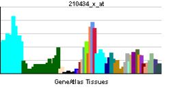
The JTB family of proteins contains several jumping translocation breakpoint proteins or JTBs. Jumping translocation (JT) is an unbalanced translocation that comprises amplified chromosomal segments jumping to various telomeres. JTB has been found to fuse with the telomeric repeats of acceptor telomeres in a case of JT. Homo sapiens JTB (hJTB) encodes a transmembrane protein that is highly conserved among divergent eukaryotic species. JT results in a hJTB truncation, which potentially produces an hJTB product devoid of the transmembrane domain. hJTB is located in a gene-rich region at 1q21, called EDC (Epidermal Differentiation Complex).[1] JTB has also been implicated in prostatic carcinomas.[3]
KANOME ET AL SUGGESTED
"JTB-induced clustering of mitochondria around the nuclear periphery and swelling of each mitochondrion. In those mitochondria, membrane potential, as monitored with a JC-1 probe, was significantly reduced. Coinciding with these changes in mitochondria, JTB retarded the growth of the cells and conferred resistance to TGF-
 1-induced
apoptosis. These activities were dependent on the N-terminal processing
and induced by wild-type JTB but not by a mutant resistant to cleavage.
These findings raised the possibility that aberration of JTB in
structure or expression induced neoplastic changes in cells through
dysfunction of mitochondria leading to deregulated cell growth and/or
death."
1-induced
apoptosis. These activities were dependent on the N-terminal processing
and induced by wild-type JTB but not by a mutant resistant to cleavage.
These findings raised the possibility that aberration of JTB in
structure or expression induced neoplastic changes in cells through
dysfunction of mitochondria leading to deregulated cell growth and/or
death."==================================================================
ONCE AGAIN YOU SEE HOW THE CELL PLAYS, USING SIMPLE THINGS THAT GET COMPLICATED REALLY QUICK!