Showing posts with label integrins. Show all posts

Friday, August 23, 2013

THE "CHELOID FACTOR" AT THE CELLULAR MEMBRANE!

We tend to be excited about intracellular pathways as they travel through the Cytosol and affect epigenetic and nuclear phenomena. And our excitement has been justified since we have been able to affect cellular life by targeting various pathway molecules.  But one should stress a particular event occurring at the membrane that mimics "wound phenomena".  Aside for providing a physical boundary of the cell, the membrane is one of the most important "organs" of the cell.  It is in itself a very chemically vibrant living "cellular tissue ".  When you start reading about the cell they tell you about the layers of proteins and lipids that make up the cellular membranes.  But this picture is far from the truth, the membrane is like the wall of a brick house.  With each brick different from the next.  Some of these bricks are called Integrins (I guess because they are an integral part of the membrane).  Some of these bricks have a Cyclin, some have a growth factor!  In fact, the membrane here serves as a reserve of these molecules. Some bricks can be divided in 2 portions.  One portion that can "FLIP" inside when needed (This portion contains the cyclin, for example) and one portion that can "FLOP" outside (this portion contains a Metalloprotease).  (see my post on FLIPPASE and FLOPPASE) The point is that once the brick is used there remains a hole with sharp edges.  These edges are called "FOCAL ADHESION Molecules" (KINASES) in a cell and are governed by the PTK2 gene!  (and of course PYK2)

PTK2:

From Wikipedia, the free encyclopedia
Jump to: navigation, search
Protein tyrosine kinase 2

PDB rendering of the C-terminal FAT domain based on 1k04[1].
Available structures
PDB Ortholog search: PDBe, RCSB
Identifiers
Symbols PTK2; FADK; FAK; FAK1; FRNK; PPP1R71; p125FAK; pp125FAK
External IDs OMIM600758 MGI95481 HomoloGene7314 ChEMBL: 2695 GeneCards: PTK2 Gene
EC number 2.7.10.2
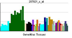
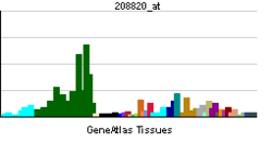
RNA expression pattern
PBB GE PTK2 207821 s at tn.png
PBB GE PTK2 208820 at tn.png
More reference expression data
Orthologs
Species Human Mouse
Entrez 5747 14083
Ensembl ENSG00000169398 ENSMUSG00000022607
UniProt Q05397 P34152
RefSeq (mRNA) NM_001199649 NM_001130409
RefSeq (protein) NP_001186578 NP_001123881
Location (UCSC) Chr 8:
141.67 – 142.01 Mb
Chr 15:
73.21 – 73.42 Mb

PubMed search [1] [2]
PTK2 protein tyrosine kinase 2 (PTK2), also known as Focal Adhesion Kinase (FAK), is a protein that, in humans, is encoded by the PTK2 gene.[2] PTK2 is a focal adhesion-associated protein kinase involved in cellular adhesion (how cells stick to each other and their surroundings) and spreading processes (how cells move around).[3] It has been shown that when FAK was blocked, breast cancer cells became less metastastic due to decreased mobility.[4](Wikepedia
=============================================================================

AND THEY ARE PLENTY TALKED ABOUT! 
===============================================================  I.E....

"Integrin-dependent translocation of phosphoinositide 3-kinase to the cytoskeleton of thrombin-activated platelets involves specific interactions of p85 alpha with actin filaments and focal adhesion kinase(JCB)"


The point is that at the membrane healing should occur after the "integrin" has been plucked off, but failure to heal may trigger the "cheloid effect".  In the cell, this is where the Src gene is, the Wnt (catenins) and the Notch are here, Caspase 3 is present, and death Receptors,etc... (things can get complicated really fast with these guys around! unless of course phosphorylation or other taming mechanisms come to play!)

Focal Adhesion kinases (FAK)

". FAK is typically located at structures known as focal adhesions, these are multi-protein structures that link the extracellular matrix (ECM) to the cytoplasmic cytoskeleton. Additional components of focal adhesions include actin, filamin, vinculin, talin, paxillin, tensin[7] and RSU-1."  This is what Taxol and Taxotere find their might!  (components of microtubules)

remember tensin is same as PTEN

NIH

" PTEN1

Also known as
BZS; DEC; CWS1; GLM2; MHAM; TEP1; MMAC1; PTEN1; 10q23del
Summary
This gene was identified as a tumor suppressor that is mutated in a large number of cancers at high frequency. The protein encoded this gene is a phosphatidylinositol-3,4,5-trisphosphate 3-phosphatase. It contains a tensin like domain as well as a catalytic domain similar to that of the dual specificity protein tyrosine phosphatases. Unlike most of the protein tyrosine phosphatases, this protein preferentially dephosphorylates phosphoinositide substrates. It negatively regulates intracellular levels of phosphatidylinositol-3,4,5-trisphosphate in cells and functions as a tumor suppressor by negatively regulating AKT/PKB signaling pathway. [provided by RefSeq, Jul 2008]"

Monday, March 11, 2013

THE CRCBM RECOMMENDS THIS PIECE OF ARTICLE TO ALL READERS!


Mechanisms of Resistance to Anti-Angiogenic Therapy and Development of Third-Generation Anti-Angiogenic Drug Candidates
  1. Peter Carmeliet
+ Author Affiliations
  1. Vesalius Research Center (VRC), Leuven, Belgium
  1. P. Carmeliet, MD, PhD, Vesalius Research Center, VIB, K.U. Leuven, Campus Gasthuisberg, Herestraat 49, B-3000, Leuven, Belgium Email: peter.carmeliet@med.kuleuven.be
  2. -----------------------------------------------------------------------------------------
  3.  
  4.  
  5. Suffice is to say that the concerns mentioned in this review, which is an excellent review, unveils in pretty good details the insufficiency of a monotherapy attacking an essential function of the cells. Not only will the cell have an answer such as dummy receptors, secondary amplification of transcription factors of growth factors, but escape mechanisms that include escape of the area leading to metastasis.  I should confess that recruiting other cells to help fight the attacker (Myeloid and endothelial cells) showed clearly how much angiogenesis is globally needed.  I would think that the reaction by the NF-kB would be sufficient; with its secondary growth  factor production, induction would be the predictable way.  But clearly, the cell wants restoration of the angiogenic function and finally wins, making Avastin effects short lived.  By inducing Hypoxia, stress becomes a secondary impetus and c-JUN enters the dance and fights again with resulting amplification of growth factor and various dislocation of various cyclins at integrin locations including the Angiopoietins.
  6. One of the things that needs to be emphasized or not looked at or discussed in your piece are events happening at the MEK.  You know by now that MEK is clearly amplified either by the cancerous process or in reaction to the blockage or consumption at VEGF.  Tracking MEK is important, because if amplified and mutated it may reverse mesengial transformation and render the cell more omnipotent.  It may be at the center of the observation that blocking both EGFR and VEGF reduces the progression free survival. Events at the MEK need to be scrutinized.
  7. You also realize that, in the long run,  MTOR will be secondarily stimulated leading to Telomere preservation (stabilization) and cell surviva
  8. The quick restoration of the angiogenic function after cessation of the treatment marks the importance of VEGF.
Your discussion has not only brought up in details the predicted failure of mono-target-therapies, but in the case of an important/critical cellular function being impaired by Avastin, and the resulting multitude of cellular responses. I really thank the authors for this important piece!
Clearly, Avastin is never meant to be a monotherapy, that is the answer!  To all action, there is a reaction. And cells expect action, it is built for them!

Sunday, February 17, 2013

TTP

THROMBOTIC THROMBOCYTOPENIC PURPURA (HYPOTHESIS CONTINUES)

Here is the example of a metastatic mechanism going bad when it hits the right ADAM.  Remember that ADAM are made of 2 basic domains, one an integrin and one a metalloproteinase domain.  The Metalloproteinase enters the Flippase-floppase (or sometimes scramblase)-like structure and is destined to be rejected outside, the Integrin is sent inside the cell.
Metalloproteinases are sent outside and attack collagen-like molecules to open the way to cell migration and allow Metastatic processes to move forward.  However, the cell membranes have a collagen-like structure, too.  So potentially the released Metalloproteinases could attack the cell.  The cell is not stupid and knows what metalloproteinase it has put out.  So it shields itself with Inhibitors and Decoy receptors from that specific metalloproteinase and the cell goes about its migration.

2 conclusions:

1. Insufficient inhibitors and decoy receptors to Metalloproteinases (and others such as Hydroeicosatetraenoic Acid) will have devastating effects.  If genetically the inhibitors are insufficient, and metalloproteinases are expressed massively, and this happens at the endothelial cell, massive and extensive destruction of endothelial cells throughout the body happens, exposing collagen like structures of the blood vessel walls.  This of course trigger extensive activation of platelets and the Thrombosis of TTP-like syndrome.  So in TTP, it is the inhibitor that is lacking.  (ie Von Willebrand cleaving protease inhibitors have also been cited).  And plasmapheresis removes the the Metalloproteinases (and microbial Antigens/toxin when relevent), stopping the onslaught.

2.What is in the Integrin domain is critical, in ADAM-17, the integrin domain is occupied by TNF-alpha, converting enzyme (TACE) which will free and activate the devastating Tumor Necrosis Factor.  Released massively, TNF can not only induce Apoptosis like certain other Cyclins (interleukine and Interferon ), but also leads directly to NECROSIS.  A massive uncontrolled septic-like syndrome kills rats after infusion of TNF.

ADAM 10, the disintegrin there gives you Amyloid structures of Alzheimer's.  TO BE SHORT, PICK THE ADAM AND SEE THE CONSEQUENT DISEASE ON YOUR OWN!

ADAM! A FLIPPASE, FLOPPASE AND/OR A SCRAMBLASE-LIKE MOLECULE !

Saturday, February 16, 2013

HYPOTHESIS : WHERE DO CYCLINS COME FROM?

There is increasing evidence that Cyclins are integrins and so are Tumor growth factors, Tumor Necrosis factors, interleukins and interferons.
All these are membrane proteins with a particularity to be released from Metalloprotease and related adhesion molecules depending on the nature of stimuli.  The discovery and description of ADAMs as type I membrane protein containing Metalloproteinase and an integrin domain locate the growth factors and Cyclins squarely at the membrane  (surface and reticulum membrane).  These proteins, once released, go straight to the Nucleus to unveil their might by activating transcription factor.  In their track to the nucleus they can amplify and activate signal transduction pathways as well as either molecules. The cyclins find cytoplasmic and protein substrates (mostly enzymes)  which have their specific domains and link to the site to activate them most of the time, changing their shapes so as to expose hidden electrons or atomic groups (such SH) to cause downstream chain activation.

Now as the pathway unfolds at light speed (or electronic speed) it may overwhelm the cell, protection has to be assured to hide death domains (which also are integrins and therefore at the membrane) and pathways to Apoptosis.  Protection at the membrane seems to be offered by the INK while the CIP/Kip.  But deep in the cell are the Bcl-like proteins. The CIP/Kip seems to work like Decoy specific proteins since the have Cyclin domain to stop them from stimulating their respective CDKs (Cyclin dependent Kinases).  Some CDKs need 2 or more different stimulations to accomplish their deed. And with the number of stimulations comes the consequent activation of various substrates.  The Retinoblastoma substrate governs the G1 progression phase in the cell cycle, but it needs at least 2 activations, first by Cyclin D followed by activation by Cyclin E in order for it to free E2F that light up tarnscrptions genes which control the path to S-phase.  This Cyclin E also activates processes leading to Histone Biosynthesis, Centrosome activity and DNA replication.  And in fact, Cyclin E is the one that leads to gene instability that characterize many triple negative breast cancers

(E2F AND CYCLIN E, ARE POWERFUL TARGETS FOR CANCER CURE)

One of the CIP/Kip(s) is the P21 which plays a role in the cell cycle arrest due to P53 activation.
I should note that the Kinase itself may be mutated.  CDK4 is mutated in Melanoma, it renders the INK4 protein unable to occupy its domain and therefore is free to affect the nuclear transcription factor.  Therefore the solution is to increase the ligand to INK4 so as to increase its ubiquitination and and degradation through the proteasome (Ipilimumab/CTLA 4 in T cell/ does not do this unfortunately, so there is more room for you to research).  YES, LIKE FOR MERCEDES, WE NEED THE E CLASS OF PROTEINS TO FURTHER UBIQUITINATION.  A MUTATION IN E CLASS (WHICH INCLUDES MDM2) WILL BE BAD IN MELANOMA!

Suffice is to show that what starts at the membrane moves quickly to the nucleus in a milli-milli second in a flash and turn the life of the host around!

It is worth noting that not only Cyclins can be blocked from entering the Nucleus where they trigger transcription factor formation, but sometimes the Decoy (Cip/Kip) is stopped from entering the nucleus and cannot stop Cyclins which have entered the nucleus: this happens in breast cancer (p27 mislocation)
-----------------------------------------------------------------------------------------
THE INTEGRINS, PRESUMED SOURCE OF CYCLINS!

Monday, February 4, 2013

CELLULAR LANGUAGE (II)

In Cellular Language I published recently that we tried to emphasize that big functions of the cell start up with an on-and-off switch.  The Tic and The Tan like in MORSE language, the 1and 0 of the computer.  While this is true, there are many other simple things at the molecular level that are just as simple, but full of physiologic and scientific implications.

1.ON and OFF switch:
--------------------------
 Events that lead to cancer are sometimes an exaggeration of a signal.  The K-RAS (there exist many RAS (es) as we discussed in differentiation) has a switch called the Sons of the Sevenless which can stay on, sending signals down the cell continuously.  Activated RAS will light on 3 signal pathways:
-MAP kinase (through RAF)--->FOS, JUN (stress),  MYC (the dangerous  leading to Burkitt)-TF
-RAL/CDC42 (important in the movement of the membrane, Metastasis)
-PI3K (leading to affect on AKT/MTOR) FOXO downstream hiding the PUMA-remember) 

Mutations at the RAS itself can also cause it to stay on, as opposed to knocking it out.
Remember, Mutations of RAS occur in 80% of Pancreatic cancers and 50% of colon cancer. 
Therefore, a simple switch can kill you with one of the most devastating diseases.

2. Change of shape:
---------------------
To confuse and look smart, your scientist calls this post-translational conformation to emphasize that this change has occurred later because of the nteraction with another molecule (in general).

At the surface of the membrane, there are here and there some Molecules called INTEGRINS; these are of various types and increasing molecular diversity and are best known as Receptors. They are large complexes of molecules gathered in chunks called 'subunits'.  They basically cross the thickness of the cellular membrane and,  outside the cell, they sense what is going on. Integrins serve many great purposes including cell division, proliferation, migration, adhesion to each either, differentiation, sensing etc. You name it, they do it!  Only division of DNA, this, they don't do.  But the membrane has to be divided also to make 2 cells in cell divison.   Even anti-coagulation happens here.  The versatility of the integrins is linked to the variety of subunits it is composed with.  Some are nature of cell specific and some are contact specific.  By contact specific, I mean what molecule outside the cell it will attach to (ie fibronectin Vs GPIIb).

Suffice is to say that kinking of the Integrins causes exposure of some parts of the Integrin molecule not naturally exposed.  If one looks at the skin of the joint at the back of the finger, one will see folded skin. if you forcefully bend your finger, the fold you were looking at will unfold and the bottom of the fold will come out.  This is what happen to the integrin when it meets outside the cell another molecule such as TALIN.

That bending triggers the attachment of other molecules or ions (phosphorylation) to the now exposed skin, lighting up the Integrins for the cascade of events which will unfold, including the activation of SRC in Sarcoma.
The shape imposed by the bending is also Molecule specific.  Despite the resemblance of SRC with the c-ABL (leukemia), their bending does not offer the same shape, and therefore, different parts of the molecule are exposed and 2 different diseases result.

CELL ADHESION- "join at the hip"
----------------------------------------
At many points the cells are joined to each other at the hips of the Talins.  Say,  at the hip of the Talin which linked to one Integrin, there is a PLUS SIGN at Cell A, and at the next TALIN attached to the Integrin of cell B, there is a NEGATIVE sign.  These 2 integrins will be attached, and cell adhesion is achieved.  Simple as that!

Wheels of cellular migration,
-----------------------------,
Cells can roll over other cells by progressively attaching Talin to Talins and breaking the talin-talins (integrin-integrin) behind, engulfing the integrins and using them again in the forthcoming attachment like a wheel touching the ground.  The cell is that smart at the membrane.

More simple things to come...
Hiding the PUMA behind the FOXO to have a death TRAP (Apoptsosis) in case the FOXO is compromised!
Just simple, but effective tricks ...