Showing posts with label floppase. Show all posts

Friday, August 23, 2013

THE "CHELOID FACTOR" AT THE CELLULAR MEMBRANE!

We tend to be excited about intracellular pathways as they travel through the Cytosol and affect epigenetic and nuclear phenomena. And our excitement has been justified since we have been able to affect cellular life by targeting various pathway molecules.  But one should stress a particular event occurring at the membrane that mimics "wound phenomena".  Aside for providing a physical boundary of the cell, the membrane is one of the most important "organs" of the cell.  It is in itself a very chemically vibrant living "cellular tissue ".  When you start reading about the cell they tell you about the layers of proteins and lipids that make up the cellular membranes.  But this picture is far from the truth, the membrane is like the wall of a brick house.  With each brick different from the next.  Some of these bricks are called Integrins (I guess because they are an integral part of the membrane).  Some of these bricks have a Cyclin, some have a growth factor!  In fact, the membrane here serves as a reserve of these molecules. Some bricks can be divided in 2 portions.  One portion that can "FLIP" inside when needed (This portion contains the cyclin, for example) and one portion that can "FLOP" outside (this portion contains a Metalloprotease).  (see my post on FLIPPASE and FLOPPASE) The point is that once the brick is used there remains a hole with sharp edges.  These edges are called "FOCAL ADHESION Molecules" (KINASES) in a cell and are governed by the PTK2 gene!  (and of course PYK2)

PTK2:

From Wikipedia, the free encyclopedia
Jump to: navigation, search
Protein tyrosine kinase 2

PDB rendering of the C-terminal FAT domain based on 1k04[1].
Available structures
PDB Ortholog search: PDBe, RCSB
Identifiers
Symbols PTK2; FADK; FAK; FAK1; FRNK; PPP1R71; p125FAK; pp125FAK
External IDs OMIM600758 MGI95481 HomoloGene7314 ChEMBL: 2695 GeneCards: PTK2 Gene
EC number 2.7.10.2
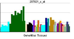
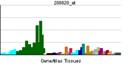
RNA expression pattern
PBB GE PTK2 207821 s at tn.png
PBB GE PTK2 208820 at tn.png
More reference expression data
Orthologs
Species Human Mouse
Entrez 5747 14083
Ensembl ENSG00000169398 ENSMUSG00000022607
UniProt Q05397 P34152
RefSeq (mRNA) NM_001199649 NM_001130409
RefSeq (protein) NP_001186578 NP_001123881
Location (UCSC) Chr 8:
141.67 – 142.01 Mb
Chr 15:
73.21 – 73.42 Mb

PubMed search [1] [2]
PTK2 protein tyrosine kinase 2 (PTK2), also known as Focal Adhesion Kinase (FAK), is a protein that, in humans, is encoded by the PTK2 gene.[2] PTK2 is a focal adhesion-associated protein kinase involved in cellular adhesion (how cells stick to each other and their surroundings) and spreading processes (how cells move around).[3] It has been shown that when FAK was blocked, breast cancer cells became less metastastic due to decreased mobility.[4](Wikepedia
=============================================================================

AND THEY ARE PLENTY TALKED ABOUT! 
===============================================================  I.E....

"Integrin-dependent translocation of phosphoinositide 3-kinase to the cytoskeleton of thrombin-activated platelets involves specific interactions of p85 alpha with actin filaments and focal adhesion kinase(JCB)"


The point is that at the membrane healing should occur after the "integrin" has been plucked off, but failure to heal may trigger the "cheloid effect".  In the cell, this is where the Src gene is, the Wnt (catenins) and the Notch are here, Caspase 3 is present, and death Receptors,etc... (things can get complicated really fast with these guys around! unless of course phosphorylation or other taming mechanisms come to play!)

Focal Adhesion kinases (FAK)

". FAK is typically located at structures known as focal adhesions, these are multi-protein structures that link the extracellular matrix (ECM) to the cytoplasmic cytoskeleton. Additional components of focal adhesions include actin, filamin, vinculin, talin, paxillin, tensin[7] and RSU-1."  This is what Taxol and Taxotere find their might!  (components of microtubules)

remember tensin is same as PTEN

NIH

" PTEN1

Also known as
BZS; DEC; CWS1; GLM2; MHAM; TEP1; MMAC1; PTEN1; 10q23del
Summary
This gene was identified as a tumor suppressor that is mutated in a large number of cancers at high frequency. The protein encoded this gene is a phosphatidylinositol-3,4,5-trisphosphate 3-phosphatase. It contains a tensin like domain as well as a catalytic domain similar to that of the dual specificity protein tyrosine phosphatases. Unlike most of the protein tyrosine phosphatases, this protein preferentially dephosphorylates phosphoinositide substrates. It negatively regulates intracellular levels of phosphatidylinositol-3,4,5-trisphosphate in cells and functions as a tumor suppressor by negatively regulating AKT/PKB signaling pathway. [provided by RefSeq, Jul 2008]"

Sunday, February 17, 2013

TTP

THROMBOTIC THROMBOCYTOPENIC PURPURA (HYPOTHESIS CONTINUES)

Here is the example of a metastatic mechanism going bad when it hits the right ADAM.  Remember that ADAM are made of 2 basic domains, one an integrin and one a metalloproteinase domain.  The Metalloproteinase enters the Flippase-floppase (or sometimes scramblase)-like structure and is destined to be rejected outside, the Integrin is sent inside the cell.
Metalloproteinases are sent outside and attack collagen-like molecules to open the way to cell migration and allow Metastatic processes to move forward.  However, the cell membranes have a collagen-like structure, too.  So potentially the released Metalloproteinases could attack the cell.  The cell is not stupid and knows what metalloproteinase it has put out.  So it shields itself with Inhibitors and Decoy receptors from that specific metalloproteinase and the cell goes about its migration.

2 conclusions:

1. Insufficient inhibitors and decoy receptors to Metalloproteinases (and others such as Hydroeicosatetraenoic Acid) will have devastating effects.  If genetically the inhibitors are insufficient, and metalloproteinases are expressed massively, and this happens at the endothelial cell, massive and extensive destruction of endothelial cells throughout the body happens, exposing collagen like structures of the blood vessel walls.  This of course trigger extensive activation of platelets and the Thrombosis of TTP-like syndrome.  So in TTP, it is the inhibitor that is lacking.  (ie Von Willebrand cleaving protease inhibitors have also been cited).  And plasmapheresis removes the the Metalloproteinases (and microbial Antigens/toxin when relevent), stopping the onslaught.

2.What is in the Integrin domain is critical, in ADAM-17, the integrin domain is occupied by TNF-alpha, converting enzyme (TACE) which will free and activate the devastating Tumor Necrosis Factor.  Released massively, TNF can not only induce Apoptosis like certain other Cyclins (interleukine and Interferon ), but also leads directly to NECROSIS.  A massive uncontrolled septic-like syndrome kills rats after infusion of TNF.

ADAM 10, the disintegrin there gives you Amyloid structures of Alzheimer's.  TO BE SHORT, PICK THE ADAM AND SEE THE CONSEQUENT DISEASE ON YOUR OWN!

ADAM! A FLIPPASE, FLOPPASE AND/OR A SCRAMBLASE-LIKE MOLECULE !

Saturday, December 15, 2012

FLIPPASE, FLOPPASE, SCRAMBLASE

One of nature's secret and ability to hide it, is by being simple.  While we expect things to be complicated and full of contorsions,  we are startled when at the end what we find is simple to understand!  One of the things we had figured out to be simple is the role of flippase and floppase, and may be the role of scramblase.   If one looks at a battery we use to power small electric equipment, one side is positive, the other is negative.  So there is a positive pole and a negative pole.  We can conclude that the battery is polarized.  The limit of a cell or one way the cell keeps what is inside of it, is by having its membrane polarized like an electrical fence.  The cell has understood that to be electrically polarized you got to have molecules in the membranes full of electrons.   And these electron-filled-molecules need to be maintained in position no matter what !  So the cell figures we need some Flippases and floppases to put things in the order above.  Meaning if the molecules we need in position A is outside the cell in position B, flip it in the right position A no matter what.  While flippase go A to B bringing these molecule inside.  Floppase goes B to A, sending molecule outside.  The Scramblase does both functions to mix things up!

This seems simple enough but wait!
This is how the cell tells the other cell "I am a dead cell, get rid of me"
Indeed, dead cells move Phosphatidyl serine, a normally internal surface molecule, to the outside of the cell, making it one of the most powerful signals to the Macrophage that this cell needs to be attacked and removed.
This disruption in lipid molecules is also linked to Bleb formation in the membrane, another powerful sign of cell death.   It is related to Caspase activity as an inducer of death, and therefore it is related to our 2nd law of nature which induces Caspases.  YOU CAN SEE HOW SIMPLE THINGS GET COMPLICATED FAST!  (This is also linked to protein Kinase activation, by the way!)
QUESTION NOW: ARE FLIPPASE AND SCRAMBLASE TARGET FOR THERAPY?  You better believe it.

We are working hard at CRBCM, but CPRIT is resisting with the help!  Please help us!

NOTE   A is inside the cell
              B is outside the Cell, in our example.