Monday, April 22, 2013

GENES INVOLVED IN APOPTOSIS (THE FIRST LAW AT WORK)

THE FIRST LAW AT WORK:
THIS IS A COMPILATION OF EXCERPTS OF RELEVANT SCIENTIFIC WORK SUPPORTING THE FACTS COVERING THE FIRST LAW
NONE OF THE WORK DETAILED HERE COME FROM CRBCM
THIS BLOG IS NOT A SCIENTIFIC SOURCE BUT HELP READER UNDERSTAND THE FUNDAMENTAL BASIS OF THE FIRST LAW  "CELL INTEND TO PROTECT INTEGRITY OF DNA AND CELL DIVISION PROCESS".  WHEN THAT PROTECTION CAN NOT BE ASSURED, CELL DEATH SHOULD ENSUE.  ONE OF THE FIRST THING THAT CANCEROUS CELLS DO IS TO FIGHT THIS LAW AND ALLOW MISTAKE TO BE TOLERATED, HERE ARE THE GENES INVOLVED IN THE FIRST LAW!  WE THANK ALL RESEARCHERS CITED BELOW FOR THEIR CONTRIBUTION TO SCIENTIFIC ADVANCE!
=======================================================================
1.NOXA

 Noxa (Latin for damage) is a pro-apoptotic member of the Bcl-2 protein family.[4] Bcl-2 family members can form hetero- or homodimers, and they act as anti- or pro-apoptotic regulators that are involved in a wide variety of cellular activities. The expression of Noxa is regulated by the tumor suppressor p53, and Noxa has been shown to be involved in p53-mediated apoptosis.

Sun et al
 Noxa is a BH3-containing mitochondrial protein that contributes to apoptosis by disrupting mitochondrial outer membrane integrity.

Proteasome inhibitor PS-341, the representative of a new class of chemotherapeutic drugs, was capable of inducing apoptosis in cisplatin-resistant SCC cells via the endoplasmic reticulum stress. PS-341 stimulated the phosphorylation of PERK and the unfolded protein response, resulting in the induction of the transcription factor ATF-4. Importantly, the Bcl-2 homology domain 3-only (BH3-only) protein Noxa was found to be strongly induced in cisplatin-resistant SCC cells by PS-341 but not by cisplatin. The knock-down of Noxa using small interference RNA significantly abolished PS-341-mediated apoptosis in SCC cells. Using eIF2α mutant mouse embryonic fibroblasts, we found that functional eIF2α played an essential role in PS-341-induced Noxa expression. Taken together, our novel findings reveal a direct link between PS-341-induced endoplasmic reticulum stress and the mitochondria-dependent apoptotic pathway and suggest that PS-341 may be utilized for overcoming cisplatin-resistance in human SCC.
---------------------------------------------------------------------------------------------------------
2.PUMA:
The p53 upregulated modulator of apoptosis (PUMA) also known as Bcl-2-binding component 3 (BBC3), is a pro-apoptotic protein, member of the Bcl-2 protein family.[
Fribley et al
Biochemical studies have shown that PUMA interacts with antiapoptotic Bcl-2 family members such as Bcl-xL, Bcl-2, Mcl-1, Bcl-w, and A1, inhibiting their interaction with the proapoptotic molecules, Bax and Bak. When the inhibition of these is lifted, they result in the translocation of Bax and activation of mitochondrial dysfunction resulting in release of mitochondrial apoptogenic proteins cytochrome c, SMAC, and apoptosis-inducing factor (AIF) leading to caspase activation and cell death.[1]
Because PUMA has high affinity for binding to Bcl-2 family members, another hypothesis is that PUMA directly activates Bax and/or Bak and through Bax multimerization triggers mitochondrial translocation and with it induces apoptosis.[6][7] Various studies have shown though, that PUMA does not rely on direct interaction with Bax/Bak to induce apoptosis.[8][9]
PUMA function is affected or absent in cancer cells
  it does not appear that genetic inactivation of PUMA is a direct target of cancer.[3  (wikipedia) Resveratrol acts to inhibit and decrease expression of antiapoptotic Bcl-2 family members while also increasing p53 expression. The combination of these two mechanisms leads to apoptosis via activation of PUMA, Noxa and other proapoptotic proteins, resulting in mitochondrial dysfunction.[48]

3.BAK
willis at al 

 "Proapoptotic Bak is sequestered by Mcl-1 and Bcl-xL, but not Bcl-2, until displaced by BH3-only proteins"

BAX
 The p53 tumor suppressor gene product can induce apoptotic cell death through an unknown mechanism..deficiency in p53 exhibit increases in Bcl-2 and decreases in Bax protein levels in several tissues this can be determined by immunohistochemical and immunoblot methods. (Miyashita et al)
Disruption of the DNA mismatch repair system, characterized by microsatellite instability (MI), plays an important role in the course of human carcinogenesis by increasing the rate of mutations of genes associated with cancers.
mutations of BAX play an important role in the course of carcinogenesis in the stomach, colorectum, and endometrium.Ouyang et al,

 Bax and Bcl-2 modulate Cdk2 activation during thymocyte apoptosis. Gil-Gomez G, Berns A, Brady HJ.

  • Up-regulation of p21WAF1 and Bax and down-regulation of Bcl-2 may be the molecular mechanism through which auristatin-PE inhibits cell growth and induces apoptosis. Li Y, Singh B, AliN,SarkarFH.
  •   pBax expression may be beneficial in predicting the effects of ACT on patients with IDC. Nio Y, Iguchi C, Yamasawa K, Sasaki S, Takamura M, Toga T, Dong M, Itakura M, Tamura K.


    Inactivation of the TGFR Beta11 appears to precede BAX Mutation which not only to block Apoptosis
    but also to  provide selective advantage for growth. (remember at this point the exasperated tumor growth factor is amplified through the stress NF-kB/c-FOS route, and act in a autocrine faction on other susceptible receptor  (Lonov et al)
-------------------------------------------------------------------------------------
4.BID
 This gene encodes a death agonist that heterodimerizes with either agonist BAX or antagonist BCL2. The encoded protein is a member of the BCL-2 family of cell death regulators. It is a mediator of mitochondrial damage induced by caspase-8 (CASP8); CASP8 cleaves this encoded protein, and the COOH-terminal part translocates to mitochondria where it triggers cytochrome c release. Multiple alternatively spliced transcript variants have been found, but the full-length nature of some variants has not been defined. [provided by RefSeq, Jul 2008]

Lee et al!
  BID, a pro-apoptotic member of the Bcl-2 family, interconnects the extrinsic apoptosis pathway initiated by death receptors to the intrinsic apoptosis pathway.

=========================================================================
5.APAF-1

This gene encodes a cytoplasmic protein that forms one of the central hubs in the apoptosis regulatory network. This protein contains (from the N terminal) a caspase recruitment domain (CARD), an ATPase domain (NB-ARC), few short helical domains and then several copies of the WD40 repeat domain. Upon binding cytochrome c and dATP, this protein forms an oligomeric apoptosome. The apoptosome binds and cleaves caspase 9 preproprotein, releasing its mature, activated form. The precise mechanism for this reaction is still debated though work published by Guy Salvesen suggests that the apoptosome may induce caspase 9 dimerization and subsequent autocatalysis.[5] Activated caspase 9 stimulates the subsequent caspase cascade that commits the cell to apoptosis.
Alternative splicing results in several transcript variants encoding different isoforms.[2]wikipedia

 APAF1 has been shown to interact with NLRP1,[7] Caspase-9,[7][8][9][10][11] APIP,[8] BCL2-like 1[10][11] and HSPA4.[12]  
Oligomeric Apaf-1 mediates the cytochrome c-dependent autocatalytic activation of pro-caspase-9 (Apaf-3), leading to the activation of caspase-3 and apoptosis. This activation requires ATP. Isoform 6 is less effective in inducing apoptosis

APOPTOSOME
BCL-2
BCL-2 like
BCLX
MCL-1
CASPASE 3 AND 7
CASPASE 9
P53
c-MYC

Sunday, April 21, 2013

ONE IMPORTANT MECHANISM OF NEOPLASTIC TRANSFORMATION COMING OUT OF OUR OBSERVATION:

ONE IMPORTANT MECHANISM OF NEOPLASTIC TRANSFORMATION COMING OUT
OF OUR OBSERVATION: THE FAILURE OF RECEPTORS

LET US TAKE THIS EXAMPLE!
---------------------------------------------------------------------------------------------

Tumor necrosis factor-alpha induces mucin hypersecretion and MUC-2 gene expression by human airway epithelial cells.

Source

Critical Care Medicine Department, Warren G. Magnuson Clinical Center, National Institutes of Health, Bethesda, Maryland 20892-1662.

Abstract

Tumor necrosis factor-alpha (TNF-alpha) is a multifunctional, pro-inflammatory cytokine that is capable of activating a diverse number of target genes within multiple cell types. Little information is known regarding the role of TNF-alpha in the regulation of human airway mucin hypersecretion and MUC-2 gene expression.

------------------------------------------------------------------------------------------

LET'S ASSUME
THAT A GROWTH FACTOR HAS 2 PRINCIPAL RECEPTORS AND 2 SECONDARY RECEPTORS
(IN THIS EXAMPLE TNF- alpha IS SAID TO HAVE  A "NUMBER OF TARGET GENES")
IF FOR SOME REASONS THE GROWTH FACTOR FAILED TO ACTIVATE ITS PRINCIPAL RECEPTORS,  2 THINGS HAPPEN:

1. THE PRINCIPAL RECEPTOR
initiates a stress like response which will involve the HP90, exacerbating the NF- kB
resulting in further activation of promoter genes and formation of MORE TNF- alpha

2. MORE TNF-alpha will exacerbate the effect on SECONDARY receptors still sensitive, leading to the activation/amplification of unwanted genes,

- Amplification of MUC-1 leads to an Adenoma and eventually Adenocarcinoma.
- Amplification of MUC-2 leads to a pseudomyxoma.
- May be MUC-7 will be lung and bladder Cancer?

Neutrophil elastase induces MUC5AC gene expression in airway epithelium via a pathway involving reactive oxygen species.

Source

Division of Pediatric Pulmonary Diseases, Duke University Medical Center, Durham, North Carolina 27710, USA.

Abstract

Neutrophil-predominant airway inflammation and mucus obstruction of the airways are major pathologic features of chronic airway diseases, including cystic fibrosis and chronic bronchitis. Neutrophils release elastase, a serine protease that impairs mucociliary clearance and stimulates goblet cell metaplasia and mucin production. We previously reported that neutrophil elastase increases expression of a major respiratory mucin gene, MUC5AC, by enhancing mRNA stability. However, the molecular mechanisms of elastase-regulated MUC5AC expression are not known. We hypothesized that reactive oxygen species, generated by elastase treatment, mediate MUC5AC gene expression.

MUC4 gene polymorphisms associate with endometriosis development and endometriosis-related infertility.

Source

Human Genetic Center, China Medical University Hospital, Taichung, Taiwan.

Abstract

BACKGROUND:

Mucin 4 (MUC4) plays an important role in protecting and lubricating the epithelial surface of reproductive tracts, but its role in the pathogenesis of endometriosis is largely unknown.

MUC3 human intestinal mucin. Analysis of gene structure, the carboxyl terminus, and a novel upstream repetitive region.

Source

Gastrointestinal Research Laboratory (151M2), Department of Veterans Affairs Medical Center, University of California, San Francisco, California 94121, USA.

Abstract

MUC3 is a large mucin glycoprotein expressed by the human intestine and gall bladder.

MUC6 - Wikipedia, the free encyclopedia

en.wikipedia.org/wiki/MUC6
Reid CJ, Harris A (1999). "Expression of the MUC 6 mucin gene in development of the human kidney and male genital ducts.".

PICK A NUMBER AND YOU WILL FIND WHERE THE CANCER WILL BE. THIS IS THE POWER OF RECEPTORS AND THE POWER OF GENE POLYMORPHISM! 

Let's play:
Full Name mucin 10, submandibular gland salivary mucin

MUC12 »  Mucin-12  (MUC-12)
Protein also known as:  Mucin-11 (MUC-11).
Gene name:  MUC12
This protein has been shown to exist at protein level

Expression

Ubiquitous, with higher expression in colon. Down-regulated in colorectal cancer as well as in the colon of patients with ulcerative colitis (UC) and Crohn's disease (CD).  
  • CuratedUniProtKB
Ubiquitous cytoplasmic expression in all tissues at variable levels. Highest expression in gastrointestinal tract. silver HPA027769; HPA023835

THIS EXERCISE CAN BE DONE WITH ALL ADENOCARCINOMAS!

DO YOU SEE PORTRAYED HERE A TARGET FOR THERAPY OR DIAGNOSIS?  
LET'S GO TO WORK!


In Summary,
Epithelial covers of Mucosa secrete a Mucine to protect it against infection and immune system exactions
and depending on localization the MUCINE is made a different family member of MUC -x  gene
this is helpful when you have a carcinoma of unknown primary, look at the MUC gene amplified

YOU reach the door of MESENCHYMALIZATION when you reach MUC-20, this is where epidermal and endothelial switch, where squamous switch to adenocarcinoma, where the MEK gene is located!

Aspirin, colorectal cancer and intreperitoneal chemotherapy

*THE NURSE HEALTH STUDY WHICH FOLLOWED CLOSE TO 200,000 INDIVIDUALS REPORTED OR SUGGESTED ASPIRIN  (325 MG ) REDUCED BY AT LEAST 23% THE RISK OF DEVELOPING COLORECTAL CANCER.  AND THIS WAS IN ADDITION TO CARDIOVASCULAR PREVENTION BENEFITS. APPARENTLY, THE INHIBITION OF CYCLO-OXYGENASE (COX 1 AND 2 ) HAS AN IMPACT ON CELL DIVISION, APOPTOSIS AND ANGIOGENESIS!

*In Colorectal cancer
BRAF mutation is of poor prognosis (10 months Vs 34.7 months for wild type BRAF), tumor is diploid with Microsatellite instability!  Give Vemurafenib!

*Intraperitoneal chemotherapy continues to show survival advantage in Ovarian cancers, despite the fact that the majority of Ovarian cancers with the relevant stage do not receive it!

CPRIT: A former congressman joined its board

CPRIT: A former congressman joined its board

PETE GEREN  a well known  former Democratic congressman from the Fort Worth counties, has been named to the CPRIT governing board.  He will bring much needed dynamism and fortitude to CPRIT.
He was nominated by the Speaker of the House of Representative Joe Straus to replace TOM LUCE who is now the new chief operating officer of the O’Donnell Foundation since April 1st, 2013.  The Dallas foundation of philanthropist Peter O’Donnell and his wife Edith, is reported to have contributed more than $600 million over the past 30 years to support scientific research and education.  Their link to CPRIT has been controversial at best!
In other related news, Jay Maguire, a lobbyist with long ties to Tobacco companies, was hired 3 months ago by the CPRIT Foundation, now called the Texas Cancer Coalition.
Observers have been proud of changes and the new direction that CPRIT has taken overall. We wish this important organization for Texas well!

Saturday, April 20, 2013

DOES HAIR COLOR CHANGE COMES FROM METHIONINE MALABSORPTION ???

DOES HAIR COLOR CHANGE COMES FROM METHIONINE MALABSORPTION ???

"The following Votrient side effects are common (occurring in greater than 30%) for patients taking Votrient:
These are less common side effects (occurring in 10-29%) for patients receiving Votrient:
This list includes common and less common side effects for those taking Votrient. Votrient's side effects that are very rare -- occurring in less than about 10 percent of patients -- are not listed here. Always inform your health care provider if you experience any unusual symptoms."

(SCOTT HAMILTON CARES )

Error of metabolism points to interesting targets to be used in T cell rearrangement strategy

Error of metabolism points to interesting targets to be used in T cell rearrangement strategy

Glut 2 ----To be looked at in Pancreatic Cancer  (No CNS involvement)
Glut 4
Gs alpha----(To be looked at in Sarcoma)
IRS 1
Biopterin Synthesis
FMR1
GNB
Adenyl Cyclase
===================================================
GENES INVOLVED IN SURVIVAL (ADAPTATION)

RAG 1
RAG 2
(TRANSPOSASE)
DNA -PK
XRCC4
XLF
ARTEMIS
CERNUNNOS
LAMBDA AND MU
VDJ
----------------------------------------------------------
RECEPTOR FOR ALPHA AND BETA CHAIN
CDR, 1-4
TCR
-------------------------------
 MGAT2
--------------------------

GENES INVOLVED IN GLYCOSYLATION

Neu5Ac
Neu 5GC
CMAH
MGAT
FUT8
CHO-STGAL
CHOK1
TRANSFERRIN

LYSOZOMAL LEVEL
GDP MANNOSE
----------------------------
PMM2
MPI
----------------------------
GENES OF MUSCLE DYSTROPHY
POMT1
POMGNT1
DYSTROGLYCAN
FKRP
------------------------------------
GALNT3 MODIFIES FGF23
COSMC FOLDING OF GLYCAN

ROLE OF TRANSFERRIN IN CANCER TREATMENT AND A MEANINGFUL POT POURRI. WE THANK ALL RESEARCHERS CITED FOR THEIR CONTRIBUTION TO SCIENCE!

"Transferrin imbalance can have serious health effects for those with low or high serum transferrin levels. A patient with an increased serum transferrin level often suffers from iron deficiency anemia.[5] A patient with decreased plasma transferrin can suffer from iron overload diseases and protein malnutrition. An absence of transferrin results from a rare genetic disorder known as atransferrinemia; a condition characterized by anemia and hemosiderosis in the heart and liver that leads to many complications, including heart failure. Most recently, transferrin and its receptor have been shown to diminish tumour cells by using the receptor to attract antibodies.[5]" WIKIPEDIA


Transferrin is also associated with the innate immune system. It is found in the mucosa and binds iron, thus creating an environment low in free iron that impedes bacterial survival in a process called iron withholding. The level of transferrin decreases in inflammation.["

Many conditions including infection and malignancy can depress transferrin levels. The transferrin is abnormally high in iron deficiency anemia.

David Goodsell
" several molecules called lactoferrin and ovotransferrin are similar to transferrin. These molecules, found in milk and egg whites respectively, also have strong binding sites for iron. However, their main function is not delivery. Instead, they serve to protect cells from bacteria. Since they mop up any free iron ions, they starve bacteria of a vital resource, slowing the growth of an infection."

Ponka et al.
" Cellular iron uptake and storage are coordinately regulated through a feedback control mechanism mediated at the post-transcriptional level by cytoplasmic factors know as iron-regulatory proteins 1 and 2. These proteins "sense" levels of iron in the transit pool and, when iron in this pool is scarce, they bind to stem-loop structures known as iron-responsive elements on the 5' untranslated region of the ferritin mRNA and 3' untranslated region of the transferrin mRNA. Such a binding inhibits translation of ferritin mRNA and stabilizes the mRNA for transferrin receptors. The opposite scenario develops when iron in the transit pool is plentiful. This remarkable regulatory mechanism prevents the expansion of a catalytically active intracellular iron pool, while maintaining sufficient concentrations of the metal for metabolic needs.

using transferrin and iron chelators are extremely complex. Improperly managed iron storage and delivery in cell culture systems is a major contributor to oxidative stress and protein damage. For a more complete discussion of transferrin and iron chelators as a cell culture components go to Sigma's Media Expert.
Back to top


Primary Functions of Transferrin in Cell Culture Systems:
  • Transferrins facilitate extracellular iron storage, and transport.
  • Transferrins are important extracellular antioxidants. They bind iron so tightly under physiological conditions that virtually no free iron exists to catalyze the production of free radicals.
  • The delivery of iron to cells by transferrins is a receptor-mediated and controlled process. Cells regulate the amount of iron they receive from the extracellular environment by varying transferrin receptor expression 
  •  Ceruloplasmin mediated oxidation of iron is accompanied by the conversion of di-oxygen into water. Ceruloplasmin is the only circulating molecule that provides controlled and rapid oxidation of ferrous to ferric iron.
Inoue et al
" We previously found that transferrin (Tf) differentially stimulated the growth of highly metastatic variant lines of murine melanoma and that these highly metastatic cells also had greater numbers of Tf receptors on their cell surfaces. In the present study we found that highly metastatic rat mammary adenocarcinoma cell lines also responded differentially to Tf in proliferation assays,"

Breast Cancer Res Treat. 1999 Aug;56(3):203-17.

Transferrin receptor overexpression enhances transferrin responsiveness and the metastatic growth of a rat mammary adenocarcinoma cell line.

Source

Department of Cancer Biology, University of Texas M.D. Anderson Cancer Center, Houston 77030, USA.

Abstract

"We previously found that breast cancer cell transferrin receptor expression and proliferative response to transferrin often correlated with metastatic capability.
 We previously found that transferrin (Tf) differentially stimulated the growth of highly metastatic variant lines of murine melanoma and that these highly metastatic cells also had greater numbers of Tf receptors on their cell surfaces. In the present study we found that highly metastatic rat mammary adenocarcinoma cell lines also responded differentially to Tf in proliferation assays,

" Nochoson et al.
The growth of brain-metastasizing melanoma cells appears to be stimulated by other paracrine growth factors, such as paracrine transferrin. Melanoma cells metastatic to brain express higher numbers of transferrin receptors and respond and proliferate at lower concentrations of transferrin than do melanoma cells metastatic to other sites or poorly metastatic melanoma cells. The results suggest that degradation and invasion of brain basement membrane and responses to paracrine neurotrophins and paracrine transferrins are important properties in brain metastasis of murine and human malignant melanoma cells."

" A major organ-derived (paracrine) growth factor has been isolated that differentially stimulates the growth of cells metastatic to the brain. Characterization of this mitogen demonstrated that it is a transferrin-like glycoprotein; cells that are metastatic to brain express greater numbers of transferrin receptors on their surfaces than cells that are poorly metastatic or metastatic to other sites. Transferrin-like factors are expressed in fetal brain and could represent the transferrin-like factors that stimulate growth of brain-"metastatic melanoma and breast cancer cells. These and other factors are probably important in determining whether metastatic cells can successfully invade, colonize, and grow in the CNS."

Reversal by Transferrin of Growth-Inhibitory Effect of Suramin on Hormone-Refractory Human Prostate Cancer Cells

  1. Warren D. W. Heston*
+ Author Affiliations
  1. W. D. W. Heston (Urologic Oncology Research Laboratory, Molecular Pharmacology and Therapeutics Section, Sloan-Kettering Institute for Cancer Research; and Urology Service, Department of Surgery, Memorial Hospital, Memorial Sloan-Kettering Cancer Center), R. S. Israeli (Urologic Oncology Research Laboratory, Molecular Pharmacology and Therapeutics Section, Sloan-Kettering Institute for Cancer Research), W. R. Fair (Urology Service, Department of Surgery, Memorial Hospital, Memorial Sloan-Kettering Cancer Center), New York, N. Y.
  1. *Correspondence to: Waren D. W. Heston, Ph.D., Memorial Sloan-Kettering Cancer Center, 1275 York Ave., Box 334, New York, NY 10021. 
" Transferrin is abundant in bone stroma and has been found to stimulate models of hormone-refractory metastatic prostate cancer. Suramin, a compound that has been used to treat metastatic prostate cancer, has been demonstrated to antagonize the binding of transferrin to the transferrin receptor and to suppress uptake of iron by hematopoietic cells. Purpose: The purpose of our study was to determine whether transferrin may"

Antisense oligonucleotide targeted to the transferrin receptor gene suppresses tumor growth and lung metastases in 4T1 mammary adenocarcinoma mouse model

X. Jiang, A. Israyeeyan, E. Boykin, L. Lomax, J. F. Head and R. L. Elliott Mastology Research Institute, Baton Rouge, LA; School of Veterinary Medical, LSU, Baton Rouge, LA
10616
Iron is required for the activity of ribonucleotide reductase and synthesis of DNA. Cellular surface transferrin receptors (TfR) are the principal transport protein for iron into cells. It has been reported that inhibition of TfR gene expression leads to the suppression of tumor cell growth in cell culture. In the present study, an antisense oligonucleotide targeted to the transferrin receptor gene was used to treat 4T1 mammary adenocarinoma in cell culture and a mouse model. The cytotoxicity of a 24-mer TfR antisense oligonucleotide was determined by 3[H]-thymidine incorporation in vitro. The sense and random sequence
BASALOID TRIPLE NEGATIVE BREAST CANCER RESULT FROM A DEFECT IN GLYCOSYLATION OF RECEPTOR PROTEIN OR A FORM OF CONGENITAL EXOSTOSIS.

We know that the disease is rare like 1/5000 women
that it occurs at age 20-40 like in Polycystic kidney disease when these things develop
it changes the morphology of the cell like dysfunction of molecular membrane would
the receptor lacking post translational  modification by glycosylation will impact their binding of any growth factor  This stresses the cell which will increase further growth factor chronically to justify SECONDARY amplification PIK/MAPK.  The stress will involve particularly the TNF receptor (exostin 1) /TRAP1.
TRAP1 (Heat stroke protein) at cellular is a stimulator of c-JUN/FOS/STAT which further increase growth factor which can effectively act on their receptors.  This persistent vicious cycle leads to the neoplastic process.
Remember when c-FOS is stimulated, c-MYC is not far away to insure proliferation is rapidly started and maintained. 
The defect however start in the Golgi apparatus where Glycosylation should have occurred
and the Wnt is involved since it influences Reticulum Endothelial contact with the Golgi Apparatus!

Yes it becomes imperative to look into EXT 1,2, Retinoblastoma 1 (Rb1), and TRAP1 gene in this disease
because genetic intervention will clearly impact thousands of women. 


This analogy at Estrogen receptor, will affect other ER negative Breast cancer
It is evident that we underestimate the importance of post translational Glycosylation of Receptors in Breast cancers. BUT IT PUT HEPARAN SULFATE METABOLIC DEFICIENCY CENTER TO THIS DISEASE!

REMEMBER THE RULE THAT WHAT CAN CHANGE THE SHAPE AND DEVELOPMENT OF A MEMBRANE OR A HUMAN EXTREMITY (OR STATURE) CAN CAUSE CANCER IF DISTORTED GENETICALLY.   AND ALWAYS REMEMBER THAT A PERSONALITY CHANGE WILL BE ASSOCIATED IN MANY CASES.   CLINICIANS TREATING POLYCYSTIC KIDNEY FORGET TO INVOLVE A PSYCHIATRIST LEAVING THEIR PATIENTS GO THROUGH BROKEN MARRIAGES, PROBLEM AT WORK AND CASES OF SUICIDES!  BE UP IN THE KNOW, GIVING ACE INHIBITORS IS NOT ENOUGH!

WHAT WALKS LIKE A DUCK, QUACKS LIKE A DUCK, LOOKS LIKE A DUCK, IS PROBABLY A DUCK, I WAS TAUGHT IN MY RESIDENCY PROGRAM !
INTRODUCTION TO THE 8TH LAW OF NATURE

THE 8TH LAW INVOLVES OF THE CELL
It is the ultimate purpose of the cell.  All cell live to survive and the cell will escape death by all costs until evidences impose a programed death as the ultimate door to escape stress.  But before its death the cell will unveil an infinite number of adaptative strategies which include resistance to new infection, autophagy, escaping immune system detection, autocrine growth factor compensation and self advantage, Activating variable genes for antibody production and T-cell receptor function, decreasing adhesion molecule, metastasizing  for cancer cells, exostosis of toxic material.  All these function are controlled by genes.  The cell has adaptation potential to even foreign threats never seen by its parents before!

The genes involved start from Transposases (RAG 1, 2) to Artemis, Cernunnos,XRCC4, XLF, VDJ, to FUT8, MGAT, CHOK1 and PMM2, MGAT2
They also include some Genes of differentiation, rearragment genes, glycosylation genes and adaptation genes, plicing and linkage genes.

full groups by class of genes and Nomenclature will follow!

TRIPLE NEGATIVE BREAST CANCER WITH BASAL CELL LIKE CANCER CELLS

Are you kidding me or just fooling me?
Could patients having this disease also have a Glycosylation problem?
Are they suffering from a form of Hereditary Multiple exostosis?
Do they have EXT gene Mutation?
 By claiming the following:
"The mechanism of HME pathology is likely rooted in a disruption of the normal distribution of HS-binding growth factors, which include FGF and morphogens such as hedgehog, Wnt, and members of the TGF-β family. The loss of HS disrupts these pathways"

Hudson Freeze et al are reporting all the mechanisms involved in Basal cell  breast cancer in one sentence and pin it down to Heparan surface.  He is putting the disruption square and round in the Golgy Apparatus.

It makes perfect sense though, these diseases are triple negative pointing to an insensitive receptor
and sure enough we know that receptors are proteins, but in order to work, proteins need Glycosylation and Heparan Sulfate is  :

"Heparan sulfate /hep·a·ran sul·fate/ (hep´ah-ran) is a glycosaminoglycan occurring in the cell membrane of most cells, consisting of a repeating disaccharide unit of glucosamine and uronic acid residues, which may be acetylated and sulfated; it accumulates in several mucopolysaccharidoses."(wikipedia)

Lack of Glycosylation will render receptor insensitive!

THIS IS THE FULL TEXT OF THEIR CLAIM:
Congenital Exostosis
Defects in the formation of heparan sulfate (HS) cause hereditary multiple exostosis (HME), an autosomal dominant disease with a prevalence of about 1:50,000. It is caused by mutations in two genes EXT1 and EXT2, which are involved in HS synthesis. HME patients have bony outgrowths, usually at the growth plates of the long bones. Normally, the growth plate contains chondrocytes in various stages of development, which are enmeshed in an ordered matrix composed of collagen-chondroitin sulfate. In HME, however, the outgrowths are often capped by disorganized cartilagenous masses with chrondrocytes in different stages of development. About 1–2% of patients also develop osteosarcoma.
HME mutations occur in EXT1 (60–70%) and EXT2 (30–40%). The proteins encoded by these genes are thought to exist as a complex in the Golgi and both are required for polymerizing GlcNAcα1–4 and GlcAβ1–3 into HS. However, the partial loss of one allele of either gene appears sufficient to cause HME. This means that haploinsufficiency decreases the amount of HS and that EXT activity is rate limiting for HS biosynthesis. This is unusual because most glycan biosynthetic enzymes are in substantial excess.
The mechanism of HME pathology is likely rooted in a disruption of the normal distribution of HS-binding growth factors, which include FGF and morphogens such as hedgehog, Wnt, and members of the TGF-β family. The loss of HS disrupts these pathways in Drosophila. Mice that are null for either Ext gene are embryonic lethal and fail to gastrulate; however, Ext heterozygous animals are viable and about one third develop a visible exostoses on the ribs. No exostoses develop on the long bones of these animals (in contrast to patients with HME), but subtle chondrocyte growth abnormalities were seen in the growth plates of these bones. Further studies are needed to understand how truncation of the HS chains leads to ectopic growth plate formation and the phenotype abnormalities.

It becomes urgent to test EXT gene in these patients the TRAP1 gene

TRAP1,
Retinoblastoma protein.[5]

EXT1

From Wikipedia, the free encyclopedia
Jump to: navigation, search
Exostosin 1
Identifiers
Symbols EXT1; EXT; LGCR; LGS; TRPS2; TTV
External IDs OMIM608177 MGI894663 HomoloGene30957 GeneCards: EXT1 Gene
EC number 2.4.1.225 2.4.1.224, 2.4.1.225
RNA expression pattern
PBB GE EXT1 201995 at tn.png
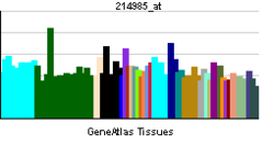
PBB GE EXT1 214985 at tn.png
More reference expression data
Orthologs
Species Human Mouse
Entrez 2131 14042
Ensembl ENSG00000182197 ENSMUSG00000061731
UniProt Q16394 P97464
RefSeq (mRNA) NM_000127 NM_010162
RefSeq (protein) NP_000118 NP_034292
Location (UCSC) Chr 8:
118.81 – 119.12 Mb
Chr 15:
53.06 – 53.35 Mb

PubMed search [1] [2]
Exostosin-1 is a protein that in humans is encoded by the EXT1 gene.[1]
This gene encodes an endoplasmic reticulum-resident type II transmembrane glycosyltransferase involved in the chain elongation step of heparan sulfate biosynthesis. Mutations in this gene cause the type I form of Multiple Exostoses.[1]
From Wikipedia, the free encyclopedia
Jump to: navigation, search
TNF receptor-associated protein 1
Identifiers
Symbols TRAP1; HSP 75; HSP75; HSP90L; TRAP-1
External IDs OMIM606219 MGI1915265 HomoloGene9457 ChEMBL: 1075132 GeneCards: TRAP1 Gene
RNA expression pattern
PBB GE TRAP1 201391 at tn.png
More reference expression data
Orthologs
Species Human Mouse
Entrez 10131 68015
Ensembl ENSG00000126602 ENSMUSG00000005981
UniProt Q12931 Q9CQN1
RefSeq (mRNA) NM_001272049 NM_026508
RefSeq (protein) NP_001258978 NP_080784
Location (UCSC) Chr 16:
3.7 – 3.77 Mb
Chr 16:
4.04 – 4.08 Mb

PubMed search [1] [2]
Heat shock protein 75 kDa, mitochondrial is a protein that in humans is encoded by the TRAP1 gene.[1][2][3]

THESE 4 GENES NEEDS TO BE LOOKED AT ASAP IN TRIPLE NEGATIVE BREAST CANCER WITH BASALOID MORPHOLOGY!

Friday, April 19, 2013

FOOD FOR THOUGHT

* A trial has shown that Doxepin rinse may reduce pains from oral Radiation induced Mucositis.

*Radiation post lumpectomy does improve survival even in the elderly
*Memantine 20 mg daily did alleviate loss of cognitive functions post whole brain Radiation
* Now that Tamoxifen can be given for 10 Years, people are still suggesting that switching at 5 years to Letrozole in postmenopausal women, is better but may be we need another clinical trial?  It is reported that switching to Letrozole lead a a 48% drop of Recurrence and 24% drop in chance of death!

*Should maintenance alpha interferon be given in Esophageal and gastric cancer with Non-mutated Interferon receptors? or cancer with over expressed NF-kB.
does overexpression of FOS predict response to Interleukin or Interferon.

*It is wrong to stop inhibitor to VEGF IN CANCER TREATMENT UNTIL DISEASE PROGRESSION, AND INSTEAD OF STOPPING, REPLACE IT BY AN MTOR INHIBITOR, THIS WILL ADD AN OPTION TO COLON CANCERS!
ESMO DID SHOW THAT UPFRONT COMBINATION OF AVASTIN AND MTOR FAILED TO IMPROVE RESULTS (renal cancers) BUT GIVING MTOR AFTER AVASTIN FAILURE IS STILL BELIEVED TO BE BETTER (PROOF OF CONCEPT.  DOES THE ESCAPE MECHANISMS TO AVASTIN OFFER AN OPPORTUNITY TO MTOR INHIBITOR FOR ACTION OR ACTIVITY? WHAT ARE THE PREDICTORS TO MTOR INHIBITORS 'ACTION?  IS TISSUE HYPOXIA SECONDARY TO ENDOTHELIAL DISTURBANCES RESULTING FROM AVASTIN A PREDISPOSITION TO MTOR INHIBITOR ACTION?

*SHOULD WE GO AHEAD AND USE NAB-PACLITAXEL AND GEMZAR IN ADJUVANT SETTING NOW?(PANCREATIC CANCER)

*ADDING ERLOTINIB TO AVASTIN AS MAINTENANCE THERAPY AFTER FIRST LINE CHEMOTHERAPY OFFERED BETTER PROGRESSION FREE SURVIVAL (1 MONTH BETTER) IN A PHASE III TRIAL PRESENTED AT ASCO 2012! (COLON CANCER)

MORE APPROVED DRUGS

*Rivaroxaban  for acute treatment and long term secondary prevention of Venous thromboembolism
*Apixaban for the prevention of stroke and systemic arterial thrombosis in patients with non valvular Atrial Fibrillation.   Apixaban 5 mg PO BID
reduce dose to 2.5 mg if patient less than 60 kg weight
                        Creatinine >1.5
                         Age >80
*Rivaroxaban approved for atrial fibrillation at 20 mg daily if GFR >50
and  15 mg if GFR between 15 and 50
No use of Riva if GFR<15

Melanoma Antigen Gene Protein MAGE-11 Regulates Androgen Receptor

*T-cell therapy, that is genetically engineered T-cells, is a valid modality of cancer treatment.  Porter et al updated their finding at the ASH describing response rate in refractory CLL and ALL.  Patients were given a lentiviral vector that expressed a chimeric antigen receptor "with specificity for the B cell Antigen CD19 paired with CD137 and CD3-zeta." (hemonc today) some of the reported results where positively dramatic.
The experience with MAGE 11 for treatment refractory melanoma did not go so well.

Melanoma Antigen Gene Protein MAGE-11 Regulates Androgen Receptor 

What you pick to attack should not be in the brain (attack to the brain by T cell is likely), and involving a "crazy gene" the like of Androgen Receptor will have many unexpected consequences.  patient died with coma and seizures!   T-cell tissue penetration is bound to occur because it is what T cell do so your task is to pick the receptor carefully!

and don't go out there and pick p300, a potent and ubiquitous transcriptional regulator, because it is Ubiquitous!  Tissue specificity as done above in Hematologic malignancies would be more appropriate!
-----------------------------------------------------------------------------------------------

NOW A BIT OF GOOD NEWS

Infusion of Autologous dendritic cell Immunotherapy  (AGS-003) given with SUNITINIB extended survival in Metastatic Renal cell cancer!   Think Immunotherapy when the going get tough we told recently... congrats to NC researcher to have proven the point!   Follow this principle in tough Myeloma cases.  May be drendritic cell infusion will work in Myeloma!

Thursday, April 18, 2013

IMPORTANT GENE FUNCTIONS WE WILL REVISIT AND CONFIRM

1. c-MYC  increases or cause progression into S-phase and DNA replication
therefore its amplification worsen proliferation and is and prognosis factor in cancer where it is involved.
But it also means you know what you get when you block this if you can.

2. Rb1 "sequester" E2F and blocks it from causing dimerization that pushes cell into S phase, the global effect of Rb1 by hiding E2F is to block the cell in G1.
amplification of E2F1 activates CPP32  and may lead to Apoptosis.
E2F5 belong to the Dream Multicomplex structure reminiscent of CBF
Rb1 is a definite member of the Crazy Genes, it interacts with many genes including the BRCA and the Androgen Receptor gene!

This stuff act directly at Histone level and its DNA.  If you want o know gene silencing here we go:


"  leading to epigenetic transcriptional repression. Controls histone H4 'Lys-20' trimethylation. Inhibits the intrinsic kinase activity of TAF1. Mediates transcriptional repression by SMARCA4/BRG1 by recruiting a histone deacetylase (HDAC) complex to the c-FOS promoter. In resting neurons, transcription of the c-FOS promoter is inhibited by BRG1-dependent recruitment of a phospho-RB1-HDAC1 repressor complex. Upon calcium influx, RB1 is dephosphorylated by calcineurin, which leads to release of the repressor complex By similarity. In case of viral infections, interactions with SV40 large T antigen, HPV E7 protein or adenovirus E1A protein induce the disassembly of RB1-E2F1 complex thereby disrupting RB1's activity. Ref.37"  (Uniprot.org)

"In addition to bladder cancer, somatic mutations in the RB1 gene are associated with many other types of cancer. For example, changes in the RB1 gene have been reported in some cases of lung cancer, breast cancer, a bone cancer known as osteosarcoma, and an aggressive form of skin cancer called melanoma. Somatic RB1 mutations have also been identified in some leukemias, which are cancers of blood-forming cells. Somatic RB1 mutations in cancer cells inactivate pRB so it can no longer regulate cell division effectively."(nih.gov)
question: What would happen if you target Calcineurin
Any time you act on the Histone, their remodeling, and RNA, you can affect tough cancers including Leukemias/sarcoma.

3.p16 inhibit CDK4 associated to Cyclin D1 (cell division)

 p16 has a trancript,  "this transcript contains an alternate open reading frame (ARF) that specifies a protein that is structurally unrelated to the products of the other variants. The ARF product functions as a stabilizer of the tumor suppressor protein p53, as it can interact with and sequester MDM2, a protein responsible for the degradation of p53.[4" wikipedia
This stuff Mutated in Melanoma and Pancreatic cancer....

"Concentrations of p16INK4a increase dramatically as tissue ages. Therefore p16INK4a could potentially be used as a blood test that measures how fast the body's tissues are aging at a molecular level.[12]"  would it explain why Pancreatic cancer is seen more in elderly?  Can we assume this mutation in elderly with pancreatic cancer?

4. P53 acts through p21WAF1

" The p21 (CIP1/WAF1) protein binds to and inhibits the activity of cyclin-CDK2 or -CDK1 complexes, and thus functions as a regulator of cell cycle progression at G1."
" p53 activates numerous microRNAs (like miR-302a, miR-302b, miR-302c, and miR-302d) that directly inhibit the p21 expression"Wikipedia
Gives us a number of targets in sarcoma where P 53 is over active

5.p14 inhibit MDM2 and free P53 to act

" p14ARF is an alternate reading frame (ARF) product of the CDKN2A locus. Both p16INK4a and p14ARF are involved in cell cycle regulation. p14ARF inhibits mdm2, thus promoting p53, which promotes p21 activation, which then binds and inactivates certain cyclin-CDK complexes, which would otherwise promote transcription of genes that would carry the cell through the G1/S checkpoint of the cell cycle. Loss of p14ARF by a homozygous mutation in the CDKN2A (INK4A) gene will lead to elevated levels in mdm2 and, therefore, loss of p53 function and cell cycle control."

LET'S GO OUT THERE FIND SARCOMA WHERE p53 IS ACTIVATED, WE KNOW WHAT TO DO NOW!


TRIPLE NEGATIVE BREAST CANCER

At the 30th Annual Miami Breast cancer conference, this puzzling group of Breast cancer emerged one more time for discussion as it continue to be challenging to investigators.  It is a complicated topic because the disease is not unique.  Triple negative Breast Cancer is a group of diseases therefore no unique genetic Mutation was brought to light as a determinant driver.  Canadian investigators reported Mutations in P53, PTEN and PI3KCA.  There was suggestion that as a group TNBC represent 16% of Breast Cancer,  And 60% have a morphology pattern of Basal like cell cancers.  There is a reported suggestion that these basal like cell cancers had a higher P53 Mutation rate and overall higher genetic variability.  How BRCA 1, 2 Mutations fit in the group was not however discussed.

The BRCA Mutations are a challenging on their own.  Although they are interpretated to represent a familial tendency to the Breast cancer, it is good to remind ourselves they belong to a group of molecules involved in DNA repair.  We know that the neoplastic process does involve destabilizing P53 function and impairing normal control mechanisms including DNA repair and immune normal mechanisms.   There is an increased push into finding a way to discern these differences more specifically.  Whether family clustering or specific Exons will be used to make the case is still being evaluated.

Given the fact that alteration in Morphology is involved raises the issue of differentiation, status of MEK is of interest to us.  This question is of Interest to us because Globally we are talking about one pathway.
RAS/RAF/MEK/MAPK/MYC/FOS/STATs.  Disturbance at the Hormone  receptors have also been looked at since these will be also ER negative.
Given Metastatic potential of the TNBR, the Wnt pathway is also of interest.

(to be continued)

Wednesday, April 17, 2013

REFRESHER NEWS!

*Quizartinib,, an investigational TKI
show promises in FLT3 AML allowing time, with some effectiveness, for stem cell transplant

*A study confirm that Hct should be maintained below 45% in Polycythemia Vera as patient above this percentage had four-fold higher incidence of thrombotic events, a study confirmed.

*Once again, and repetition is good, Ibrutinib, a Bruton's TKI show appreciable activity in CLL alone or in combination with Rituxan.

* From Smita Bathia, MD
we learned that "genes involved in the metabolism of Anthracyclines include CBR1, CBR3,NQO1, MRP1,2
defense for Oxidative stress that can cause damage to heart cells, NCF4 RAC2 CYBA,SOD
Blood pressure and heart rate regulation AGT, AGTR1, ACE, ADRB1
Iron Homeostasis HFE"

This will prompt to plan a discussion of gene Nomenclature relevant to these genes!

*Adding Vorinostat to Chemotherapy during transplantation process reduces the incidence of Acute GVHD.
*Just a rminder again, Alterations in CYP2D6 affects efficacy of Tamoxifen.

CARNITINE
"In some of the studies, L-carnitine therapy was subsequently given for six months to a year, suggesting a possible role not only in acute management but also secondary prevention, according to the authors, led by Dr James J DiNicolantonio (Wegmans Pharmacy, Ithaca, NY)
The apparent survival gains from L-carnitine, which were only modestly significant, seemed to come from infarct-size limitation and cardiomyocyte membrane stabilization, with chronic improvements in cellular energy metabolism, according to their report published online today in the Mayo Clinic Proceedings."
THE ATTRACTION HERE IS ITS RELATION TO THE MTOR
THEY BOTH ACT ON GLUCOSE METABOLISM!

AVANDIA

"Rosiglitazone is a diabetes drug from the thiazolidinedione class; in September 2010, it was suspended by the European Medicines Agency (EMA) because it deemed that the product's cardiovascular risk outweighed its benefits.
At the same time, the FDA deemed there was "a signal of harm" but decided not to suspend the drug. Instead it requested readjudication of the Rosiglitazone Evaluated for Cardiovascular Outcomes and Regulation of Glycemia in Diabetes (RECORD) study at the individual patient-data level. The agency also required that rosiglitazone-containing medicines be subject to a risk evaluation and mitigation strategy (REMS) program to ensure the safe use of the medicine in patients with no other options. (MEDSCAPE)

was avandia going to be pulled from the market? that was the 12 millions question in the article...
don't kill the messenger!
YOU WHO GOT A LAB  (THE CRBCM CAN'T, CRBCM DOES NOT HAVE  POLITICAL BACK BONE, WE ARE MUZZLED, POLITICALLY STOPPED FOR DOING RESEARCH, SO WE GIVE YOU THE CUES)
GO GET THE CURE FOR US!

USE shRNA TO SILENCE PI3K IN MELANOMA
OR RESTORE PTEN FUNCTION!

Small hairpin RNA

From Wikipedia, the free encyclopedia
Jump to: navigation, search

Lentiviral delivery of shRNA and the mechanism of RNA interference in mammalian cells.
A small hairpin RNA or short hairpin RNA (shRNA) is a sequence of RNA that makes a tight hairpin turn that can be used to silence target gene expression via RNA interference (RNAi). Expression of shRNA in cells is typically accomplished by delivery of plasmids or through viral or bacterial vectors. The promoter choice is essential to achieve robust shRNA expression. At first, polymerase III promoters such as U6 and H1 were used; however, these promoters lack spatial and temporal control.[1] As such, there has been a shift to using polymerase II promoters to regulate expression of shRNA. shRNA is an advantageous mediator of RNAi in that it has a relatively low rate of degradation and turnover. However, shRNA is disadvantageous in that it requires use of an expression vector which can pose safety concerns.
IMPORTANT GENE INTERACTIONS II
-----------------------------------------------
2.
*In order to control Triple Negative Breast cancer, you got to control the Wnt pathway!!!
Loss of PTEN desactivates BRAFeffect by abrogating BRAF induced oncogen driven Senescence
of cancer cells.
* ACTIVATION OF BRAF INDUCES BENIGN OR GOOD PROGNOSIS LESIONS, HOWEVER MELANOMA SUPPOSE ASSOCIATED ALTERATIONS IN THE PTEN AND THEREFORE THE PI3K SIGNALING PATHWAYS OPENING THE DOOR TO A ROLE FOR THE MTOR IN MELANOMA..

*NON INHIBITION OF THE MTOR LEAD TO SUPPRESSION OF THE p15INK 48 WHICH INCREASE TELOMERE STABILITY!

IF THIS IS TRUE, PROTECTION OR RESTORATION (activation) OF PTEN SHOULD PROTECT OR PREVENT MELANOMA!

--------------------------------------------------------------
SUPPORTIVE EVIDENCES


2.1 

PTEN Loss Confers BRAF Inhibitor Resistance to Melanoma Cells through the Suppression of BIM Expression

Kim H.T. Paraiso1 et al.

"PI3K inhibitor enhanced BIM expression at both the mRNA and protein level and increased the level of apoptosis through a mechanism involving AKT3 and the activation of FOXO3a."

2.2  " Upon induction of BRaf(V600E) expression, mice developed benign melanocytic hyperplasias that failed to progress to melanoma over 15-20 months. By contrast, expression of BRaf(V600E) combined with Pten tumor suppressor gene silencing elicited development of melanoma with 100% penetrance, short latency and with metastases observed in lymph nodes and lungs. Melanoma was prevented by inhibitors of mTorc1 (rapamycin) or MEK1/2 (PD325901) but, upon cessation of drug administration, mice developed melanoma,"   Dankort et al.

3."β-catenin levels control tumor differentiation and regulate both MAPK/Erk and PI3K/Akt signaling. Highly metastatic tumors with β-catenin stabilization are very similar to a subset of human melanomas. Together these findings establish Wnt signaling as a metastasis regulator in melanoma"  Damsky et al.
Advertisement
  • "β- catenin loss in Pten/Braf melanomas improves survival and inhibits metastasis
  • β-catenin stabilization in Pten/Braf melanomas enhances metastasis
  • Highly differentiated melanomas can be very metastatic in vivo
  • β-catenin status in melanoma regulates PI3K/Akt and MAPK/Erk signaling"
2.4  " It has been reported that oncogenic activations of intracellular signaling pathways downstream of EGFR, including the RAS-RAF-MAPK and PI3K-PTEN-AKT signaling pathways, are important mechanisms for generating resistance to anti-EGFR MoAbs. In the RAS-RAF-MAPK pathway, active mutations of KRAS or BRAF are not uncommon"  Zhou et al
"The loss of PTEN expression, which was reported to occur in 19.0%–42.0% of Western and 30.0%–64.0% of Chinese CRC tumors, induces an increase in PIP-3 concentration and PIK3CA pathway activation"

2.5 "Beta-catenin Controls Metastasis in Braf-activated Pten-inactivated Melanomas" "beta-catenin levels control tumor differentiation and regulate both MAPK/Erk and PI3K/Akt signaling."


see the article.

2.6.

Abrogation of BRAFV600E-induced senescence by PI3K pathway activation contributes to melanomagenesis PTEN depletion abrogates BRAFV600E-induced senescence in human fibroblasts and melanocytes."

Liesbeth C.W. ET AL."

The PI3K pathway was often activated through either decreased PTEN or increased AKT3 expression in melanomas relative to their adjacent nevi. Pharmacologic PI3K inhibition in melanoma cells suppressed proliferation and induced the senescence-associated tumor suppressor p15INK4B. This treatment also eliminated subpopulations resistant to targeted BRAFV600E inhibition."

2.7


ICAM-1 Has a Critical Role in the Regulation of Metastatic Melanoma Tumor Susceptibility to CTL Lysis by Interfering with PI3K/AKT Pathway

  "More importantly, we found that the level of ICAM-1 expression by melanoma cells correlated with decreased PTEN activity. ICAM-1 knockdown in T1 cells resulted in increased phosphorylation of PTEN and the subsequent activation of AKT. We have additionally shown that inhibition of the phosphatidylinositol (3,4,5)-triphosphate kinase (PI3K)/AKT pathway by the specific inhibitor wortmannin induced a significant potentiation of susceptibility of G1 and ICAM-1 small interfering RNA–treated T1 cells to CTL-induced lysis. The present study shows that a shift in ICAM-1 expression, which was associated with an activation of the PI3K/AKT pathway, can be used by metastatic melanoma cells to escape CTL-mediated killing."

(PLEASE NOTE MART-1 AND ITS ROLE OR INTERACTION WITH PTEN!)