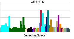
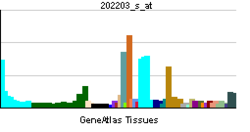
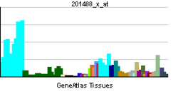
1.NMYC interactor (NMI) interacts with NMYC and CMYC (two members of the oncogene Myc family), and other transcription factors containing a Zip, HLH, or HLH-Zip motif. The NMI protein also interacts with all STATs except STAT2 and augments STAT-mediated transcription in response to cytokines IL-2 and IFN-gamma. The NMI mRNA has low expression levels in all human fetal and adult tissues tested except brain and has high expression in cancer cell line-myeloid leukemias.[3]
THIS ONE GO STRAIGHT TO LEUKEMIA AND LYMPHOBLASTIC LYMPHOMA (BURKITT)
----------------------------------------------------------------------------------------------------------
-------------------
2.Crk-like protein is a protein that in humans is encoded by the CRKL gene.[1][2]
v-CRK avian sarcoma virus CT10-homolog-like contains one SH2 domain and two SH3 domains. CRKL has been shown to activate the RAS and JUN kinase signaling pathways and transform fibroblasts in a RAS-dependent fashion. It is a substrate of the BCR-ABL tyrosine kinase and plays a role in fibroblast transformation by BCR-ABL. In addition, CRKL has oncogenic potential.[3]
CrkL together with Crk participates in the Reelin signaling cascade downstream of DAB1.[4][5]
REELIN PATHWAY, NOT VERY MUCH TALK ABOUT!
WHEN A GENE PLAYS A ROLE IN SOME KIND OF TRANSFORMATION THAT IS MORPHOLOGICALLY MEANINGFUL, IT IS AN IMPORTANT GENE!
--------------------------------------------------------------------------------------------------------------------------------------
Retinoic Acid Inhibits Serum-stimulated Activator Protein-1
Gene Expressions during
the Vitamin-induced DifferenTIATION (GO TO ARTICLE)
-------------------------------------------------------------------------------
3.Alzheimer's Disease
10 SNPs of GAB2 have been associated with late-onset Alzheimer's disease (LOAD).[14] However, this association is found only in APOE ε4 carriers.[15] In LOAD brains, GAB2 is overexpressed in neurons, tangle-bearing neurons, and dystrophic neuritis.[8][15]GAB2 has been indicated in playing a role in the pathogenesis of Alzheimer's disease via its interaction with tau and amyloid precursor proteins.[8] GAB2 may prevent neuronal tangle formation characteristic of LOAD by reducing phosphorylation of tau protein via the activation of the PI3K signaling pathway, which activates Akt. Akt inactivates Gsk3, which is responsible for tau phosphorylation.[8] Mutations in GAB2 could affect Gsk3-dependent phosphorylation of tau and the formation of neurofibrillary tangles.[8][15][16] Interactions between GAB2-Grb2 and APP are enhanced in AD brains, suggesting an involvement of this coupling in the neuropathogenesis of AD.[8]
Cancer
GAB2 has been linked to the oncogenesis of many cancers including colon, gastric, breast, and ovarian cancer.[5][12] Studies suggest that GAB2 is used to amplify the signal of many RTKs implicated in breast cancer development and progression.[4]GAB2 has been particularly characterized for its role in leukemia. In chronic myelogenous leukemia (CML), GAB2 interacts with the Bcr-Abl complex and is instrumental in maintaining the oncogenic properties of the complex.[5][12][17] The Grb2/GAB2 complex is recruited to phosphorylated Y177 of the Bcr-Abl complex leading to Bcr-Abl-mediated transformation and leukemogenesis.[4] GAB2 also plays a role in juvenile myelomonocytic leukemia (JMML). Studies have shown the protein’s involvement in the disease via the Ras pathway.[12] In addition, GAB2 appears to play an important role in PTPN11 mutations associated with JMML.[12]
GAB2, DON'T SAY I DID NOT MENTION PTPN11 AS IMPORTANT
----------------------------------------------------------------------------------------------------
4. In immunohistochemistry, CD31" is used primarily to demonstrate the presence of endothelial cells in histological tissue sections. This can help to evaluate the degree of tumour angiogenesis, which can imply a rapidly growing tumour. Malignant endothelial cells also commonly retain the antigen, so that CD31 immunohistochemistry can also be used to demonstrate both angiomas and angiosarcomas. It can also be demonstrated in small lymphocytic and lymphoblastic lymphomas, although more specific markers are available for these conditions.[7]"
FOR THOSE WHO LIKE TO QUANTIFY EVERYTHING
HOW MUCH A TUMOR HAS CD31 COULD PREDICT RESPONSE TO AVASTIN?
---------------------------------------------------------
LAIR1 prevent lysis of cells recognized as self
THIS GENE'S MUTATION NOT GOOD FOR YOU, AUTOIMUNE DISEASE LURKING
--------------------------------------------------
5.PRKCI
From Wikipedia, the free encyclopedia
| Protein kinase C, iota | |||||||||||
|---|---|---|---|---|---|---|---|---|---|---|---|
 PDB rendering based on 1vd2. |
|||||||||||
|
|||||||||||
| Identifiers | |||||||||||
| Symbols | PRKCI; DXS1179E; PKCI; nPKC-iota | ||||||||||
| External IDs | OMIM: 600539 MGI: 99260 HomoloGene: 37667 ChEMBL: 2598 GeneCards: PRKCI Gene | ||||||||||
| EC number | 2.7.11.13 | ||||||||||
|
|||||||||||
| RNA expression pattern | |||||||||||
 |
|||||||||||
 |
|||||||||||
 |
|||||||||||
| More reference expression data | |||||||||||
| Orthologs | |||||||||||
| Species | Human | Mouse | |||||||||
| Entrez | 5584 | 18759 | |||||||||
| Ensembl | ENSG00000163558 | ENSMUSG00000037643 | |||||||||
| UniProt | P41743 | Q62074 | |||||||||
| RefSeq (mRNA) | NM_002740 | NM_008857 | |||||||||
| RefSeq (protein) | NP_002731 | NP_032883 | |||||||||
| Location (UCSC) | Chr 3: 169.94 – 170.02 Mb |
Chr 3: 31 – 31.05 Mb |
|||||||||
| PubMed search | [1] | [2] | |||||||||
This gene encodes a member of the protein kinase C (PKC) family of serine/threonine protein kinases. The PKC family comprises at least eight members, which are differentially expressed and are involved in a wide variety of cellular processes. This protein kinase is calcium-independent and phospholipid-dependent. It is not activated by phorbolesters or diacylglycerol. This kinase can be recruited to vesicle tubular clusters (VTCs) by direct interaction with the small GTPase RAB2, where this kinase phosphorylates glyceraldehyde-3-phosphate dehydrogenase (GAPD/GAPDH) and plays a role in microtubule dynamics in the early secretory pathway. This kinase is found to be necessary for BCL-ABL-mediated resistance to drug-induced apoptosis and therefore protects leukemia cells against drug-induced apoptosis. There is a single exon pseudogene mapped on chromosome X.[3]
THIS IS TO LOOK AT DEFINITELY IN ALZHEIMER TOO !
RAISES A GOOD QUESTION, WHAT IS THE ROLE OF PSEUDOGENE?
---------------------------------------------------------------------------------------------------------------
6.Valosin-containing protein (VCP) is a member of a family that includes putative ATP-binding proteins involved in vesicle transport and fusion, 26S proteasome function, and assembly of peroxisomes. VCP, as a structural protein, is associated with clathrin, and heat-shock protein Hsc70, to form a complex. VCP has been implicated in a number of cellular events that are regulated during mitosis, including homotypic membrane fusion, spindle pole body function, and ubiquitin-dependent protein degradation.[3]
FOR THOSE WHO LIKED THE LYSOZOME BLOG, HERE IS YOUR GENE
ALL FUNCTION SENT TO EXTRA CELLULAR SPACE THROUGH EXOCYTOSIS CAN BE CHALLENGED THROUGH THIS GENE I PRESUME, IMPORTANT STUFF
----------------------------------------------------------------------------------------------------------------------------------------------------
7.AMFR
From Wikipedia, the free encyclopedia
| Autocrine motility factor receptor, E3 ubiquitin protein ligase | |||||||||||
|---|---|---|---|---|---|---|---|---|---|---|---|
 Rendering based on PDB 2EJS. |
|||||||||||
|
|||||||||||
| Identifiers | |||||||||||
| Symbols | AMFR; GP78; RNF45 | ||||||||||
| External IDs | OMIM: 603243 MGI: 1345634 HomoloGene: 888 GeneCards: AMFR Gene | ||||||||||
| EC number | 6.3.2.19 | ||||||||||
|
|||||||||||
| RNA expression pattern | |||||||||||
 |
|||||||||||
 |
|||||||||||
| More reference expression data | |||||||||||
| Orthologs | |||||||||||
| Species | Human | Mouse | |||||||||
| Entrez | 267 | 23802 | |||||||||
| Ensembl | ENSG00000159461 | ENSMUSG00000031751 | |||||||||
| UniProt | Q9UKV5 | Q9R049 | |||||||||
| RefSeq (mRNA) | NM_001144 | NM_011787 | |||||||||
| RefSeq (protein) | NP_001135 | NP_035917 | |||||||||
| Location (UCSC) | Chr 16: 56.4 – 56.46 Mb |
Chr 8: 93.97 – 94.01 Mb |
|||||||||
| PubMed search | [1] | [2] | |||||||||
Autocrine motility factor is a tumor motility-stimulating protein secreted by tumor cells. The protein encoded by this gene is a glycosylated transmembrane protein and a receptor for autocrine motility factor. The receptor, which shows some sequence similarity to tumor protein p53, is localized to the leading and trailing edges of carcinoma cells.[2]"
CANCER DRIVEN BY AUTOCRINE MECHANISMS COULD BE AFFECTED
--------------------------------------------------------------------------------------------------------------------------
8.There is one other NGF receptor besides TrkA, called the "LNGFR" (for "Low Affinity Nerve Growth Factor Receptor"). As opposed to TrkA, the LNGFR plays a somewhat less clear role in NGF biology. Some researchers have shown the LNGFR binds and serves as a "sink" for neurotrophins. Cells which express both the LNGFR and the Trk receptors might therefore have a greater activity – since they have a higher "microconcentration" of the neurotrophin. It has also been shown, however, that in the absence of a co-expressed TrkA, the LNGFR may signal a cell to die via apoptosis – so therefore cells expressing the LNGFR in the absence of Trk receptors may die rather than live in the presence of a neurotrophin."
WATCH THIS ONE, ANTIBODY TO Trka COULD UNLEASH APOPTOSIS THROUGH LNGFR IN THOSE CELLS HAVING BOTH GENES!
-----------------------------------------------------------------------------------------
9.The Nck (non-catalytic region of tyrosine kinase adaptor protein 1) belongs to the adaptor family of proteins. The nck gene was initially isolated from a human melanoma cDNA library using a monoclonal antibody produced against the human melanoma-associated antigen. The Nck family has two known members in human cells (Nck-1/Nckalpha and NcK2/NcKbeta), two in mouse cells (mNckalpha and mNckbeta/Grb4) and one in drosophila (Dock means dreadlocks-ortholog).
The two murine gene products exhibit 68% amino acid identity to one another, with most of the sequence variation being located to the linker regions between the SH3 and SH2 domains, and are 96% identical to their human counterparts. While human nck-1 gene has been localised to the 3q21 locus of chromosome 3, the nck-2 gene can be found on chromosome 2 at the 2q12 locus.
THIS ONE IS A TRUE CRITICAL GENE, CHECK OUT ITS INTERACTIONS!
----------------------------------------------------------------------
10. KHDRBS1
From Wikipedia, the free encyclopedia
| KH domain containing, RNA binding, signal transduction associated 1 | |||||||||||
|---|---|---|---|---|---|---|---|---|---|---|---|
|
|||||||||||
| Identifiers | |||||||||||
| Symbols | KHDRBS1; Sam68; p62; p68 | ||||||||||
| External IDs | OMIM: 602489 MGI: 893579 HomoloGene: 4781 ChEMBL: 1795190 GeneCards: KHDRBS1 Gene | ||||||||||
|
|||||||||||
| RNA expression pattern | |||||||||||
 |
|||||||||||
 |
|||||||||||
| More reference expression data | |||||||||||
| Orthologs | |||||||||||
| Species | Human | Mouse | |||||||||
| Entrez | 10657 | 20218 | |||||||||
| Ensembl | ENSG00000121774 | ENSMUSG00000028790 | |||||||||
| UniProt | Q07666 | Q60749 | |||||||||
| RefSeq (mRNA) | NM_001271878 | NM_011317 | |||||||||
| RefSeq (protein) | NP_001258807 | NP_035447 | |||||||||
| Location (UCSC) | Chr 1: 32.48 – 32.53 Mb |
Chr 4: 129.7 – 129.74 Mb |
|||||||||
| PubMed search | [1] | [2] | |||||||||
This gene encodes a member of the K homology domain-containing, RNA-binding, signal transduction-associated protein family. The encoded protein appears to have many functions and may be involved in a variety of cellular processes, including alternative splicing, cell cycle regulation, RNA 3'-end formation, tumorigenesis, and regulation of human immunodeficiency virus gene expression.[3]
---------------
DISCUSSED
No comments:
Post a Comment